0
views
0
recommends
+1 Recommend
0 collections
    0
    shares
      • Record: found
      • Abstract: found
      • Article: found
      Is Open Access

      Tension Pneumocephalus Caused by Ethmoidal Roof Fracture: Emergent Surgical Decompression

      , , ,
      Diagnostics
      MDPI AG

      Read this article at

      Bookmark
          There is no author summary for this article yet. Authors can add summaries to their articles on ScienceOpen to make them more accessible to a non-specialist audience.

          Abstract

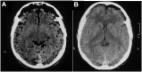
          Tension pneumocephalus is a neurosurgical emergency that occurs when air is trapped in the intracranial cavity, leading to brain compression and causing severe neurological symptoms such as decreases in motor function, sensory function, and consciousness. Most cases of pneumocephalus require conservative treatment; however, because of the possible fatal complications, rapid diagnosis and appropriate treatment are important. Here, we present the case of an 81-year-old male patient who had undergone head trauma three hours prior to being admitted to our emergency room (ER) because of mental cloudiness. The radiologic findings showed tension pneumocephalus caused by an ethmoidal roof fracture. Emergency reconstruction of the ethmoidal roof with craniotomy was performed to remove the intracranial air using normal saline irrigation. By sharing our experience with this case, we hope to provide an option for the treatment of such cases.

          Related collections

          Most cited references8

          • Record: found
          • Abstract: not found
          • Article: not found

          The Mount Fuji sign.

            Bookmark
            • Record: found
            • Abstract: found
            • Article: found
            Is Open Access

            Effects of hyperbaric oxygenation therapy on symptomatic pneumocephalus

            Background Pneumocephalus (PNC) is defined as a pathological collection of gas within the cranial cavity. The authors studied the effects of hyperbaric oxygenation (HBO2) therapy on a group of patients with PNC, comparing them with a control group to determine the relative impact on pneumocephalus volume, clinical symptoms, and duration of hospitalization. Methods Twenty-four patients with PNC treated at our hospital were consecutively studied. These patients were divided into a treated group (n=13) and a control group (n=11). Thirteen patients (treated group) were treated with HBO2 therapy sessions in a monoplace hyperbaric chamber at 2.5 atmospheres with 100% oxygen concentration. The control group was treated with normobaric oxygenation. Results Clinical improvement was seen in all patients. In the treated group, a decrease of the gas bubble was observed on the computerized tomography scan after each session of HBO2. The treated group also experienced a lower rate of meningitis compared with the control group. The length of hospital stay was significantly higher in the control group compared with the treated group. Conclusions HBO2 therapy in selective cases may lead to clinical and radiological improvement in patients with PNC.
              Bookmark
              • Record: found
              • Abstract: found
              • Article: found
              Is Open Access

              Posttraumatic delayed tension pneumocephalus: Rare case with review of literature

              Pneumocephalus is commonly seen after head and facial trauma, ear infections, and tumors of the skull base or neurosurgical interventions. In tension pneumocephalus, the continuous accumulation of intracranial air is thought to be caused by a “ball valve” mechanism. In turn, this may lead to a mass effect on the brain, with subsequent neurological deterioration and signs of herniation. Tension pneumocephalus is considered a life-threatening, neurosurgical emergency burr-hole evacuation was performed and he experienced a full recovery. However, more invasive surgery was needed to resolve the condition. Delayed tension pneumocephalus is extremely rare and considered a neurosurgical emergency. Pneumocephalus is a complication of head injury in 3.9–9.7% of the cases. The accumulation of intracranial air can be acute (<72 h) or delayed (≥72 h). When intracranial air causes intracranial hypertension and has a mass effect with neurological deterioration, it is called tension pneumocephalus. We represent a clinical case of a 30-year-old male patient with involved in a road traffic accident, complicated by tension pneumocephalus and cerebrospinal fluid rhinorrhea on 1 month after trauma and underwent urgent surgical intervention. Burr-hole placement in the right frontal region, evacuation of tension pneumocephalus. Tension pneumocephalus is a life-threatening neurosurgical emergency case, which needs to undergo immediate surgical intervention.
                Bookmark

                Author and article information

                Contributors
                Journal
                DIAGC9
                Diagnostics
                Diagnostics
                MDPI AG
                2075-4418
                January 2023
                December 28 2022
                : 13
                : 1
                : 92
                Article
                10.3390/diagnostics13010092
                ea09e633-7301-4e73-b27a-602f7d1efac2
                © 2022

                https://creativecommons.org/licenses/by/4.0/

                History

                Comments

                Comment on this article