17
views
0
recommends
+1 Recommend
0 collections
    0
    shares
      • Record: found
      • Abstract: found
      • Article: found
      Is Open Access

      Effects of distinct drugs on gene transcription in an osteosarcoma cell line

      research-article

      Read this article at

      Bookmark
          There is no author summary for this article yet. Authors can add summaries to their articles on ScienceOpen to make them more accessible to a non-specialist audience.

          Abstract

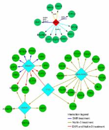
          Osteosarcoma (OS) is a common cancerous bone tumor which has a detrimental impact on the lives of patients and their families. The present study aimed at investigating the underlying molecular mechanism of various drug treatments pertaining to OS, including dimethyl sulfoxide (DMSO), doxorubicin (DXP), Nutlin-3, actinomycin D (ActD) and etoposide (Eto). Microarray and p53 chromatin immunoprecipitation combined with sequencing (ChIP-seq) datasets of the OS cell line U2OS treated with distinct drugs were acquired from the Gene Expression Omnibus and differentially-expressed genes (DEGs) were screened for alignment analysis. The p53-binding target genes were identified and ChIP-seq and microarray gene expression data were combined to identify directly and indirectly targeted genes. A regulatory network of p53 was constructed with the acquired data. Finally, the Database for Annotation, Visualization and Integrated Discovery was interrogated for annotation of target genes. A total of 212 p53-binding peaks were obtained in the untreated group, whereas thousands of peaks were obtained in the treated groups. In total, ~1,000 target genes were identified in each of DXP, DMSO, Eto and ActD treatment groups, whereas the Nutlin-3 treatment group identified an increased number, with 5,458 target genes obtained. Several common DEGs including MDM2, TP53I3, RRM2B, FAS and SESN1 were targeted by all the drugs with the exception of DMSO. p53 regulated various genes including EHF, HOXA10 and BHLHE40 in the Nutlin-3 treatment group, whereas p53 regulated EHF, RFX3, TRAF40 and TCF7L2 in the DXR treatment group. The results of the present study indicate that p53 was able to directly regulate target genes including MDM2, TP53I3 and RRM2B or indirectly regulate numerous further genes through several hub genes including EHF and RFX through various drug treatments in U2OS cells. Furthermore, p53 regulated distinct molecular processes in various drug treatments.

          Related collections

          Most cited references28

          • Record: found
          • Abstract: found
          • Article: not found

          Role of Bcl-2 family proteins in a non-apoptotic programmed cell death dependent on autophagy genes.

          Programmed cell death can be divided into several categories including type I (apoptosis) and type II (autophagic death). The Bcl-2 family of proteins are well-characterized regulators of apoptosis, and the multidomain pro-apoptotic members of this family, such as Bax and Bak, act as a mitochondrial gateway where a variety of apoptotic signals converge. Although embryonic fibroblasts from Bax/Bak double knockout mice are resistant to apoptosis, we found that these cells still underwent a non-apoptotic death after death stimulation. Electron microscopic and biochemical studies revealed that double knockout cell death was associated with autophagosomes/autolysosomes. This non-apoptotic death of double knockout cells was suppressed by inhibitors of autophagy, including 3-methyl adenine, was dependent on autophagic proteins APG5 and Beclin 1 (capable of binding to Bcl-2/Bcl-x(L)), and was also modulated by Bcl-x(L). These results indicate that the Bcl-2 family of proteins not only regulates apoptosis, but also controls non-apoptotic programmed cell death that depends on the autophagy genes.
            Bookmark
            • Record: found
            • Abstract: found
            • Article: not found

            Multiplexed massively parallel SELEX for characterization of human transcription factor binding specificities.

            The genetic code-the binding specificity of all transfer-RNAs--defines how protein primary structure is determined by DNA sequence. DNA also dictates when and where proteins are expressed, and this information is encoded in a pattern of specific sequence motifs that are recognized by transcription factors. However, the DNA-binding specificity is only known for a small fraction of the approximately 1400 human transcription factors (TFs). We describe here a high-throughput method for analyzing transcription factor binding specificity that is based on systematic evolution of ligands by exponential enrichment (SELEX) and massively parallel sequencing. The method is optimized for analysis of large numbers of TFs in parallel through the use of affinity-tagged proteins, barcoded selection oligonucleotides, and multiplexed sequencing. Data are analyzed by a new bioinformatic platform that uses the hundreds of thousands of sequencing reads obtained to control the quality of the experiments and to generate binding motifs for the TFs. The described technology allows higher throughput and identification of much longer binding profiles than current microarray-based methods. In addition, as our method is based on proteins expressed in mammalian cells, it can also be used to characterize DNA-binding preferences of full-length proteins or proteins requiring post-translational modifications. We validate the method by determining binding specificities of 14 different classes of TFs and by confirming the specificities for NFATC1 and RFX3 using ChIP-seq. Our results reveal unexpected dimeric modes of binding for several factors that were thought to preferentially bind DNA as monomers.
              Bookmark
              • Record: found
              • Abstract: found
              • Article: not found

              Actinomycin and DNA transcription.

              Recent advances in understanding how actinomycin binds to DNA have suggested its mechanism of action. Actinomycin binds to a premelted DNA conformation present within the transcriptional complex. This immobilizes the complex, interfering with the elongation of growing RNA chains. The model has a number of implications for understanding RNA synthesis.
                Bookmark

                Author and article information

                Journal
                Oncol Lett
                Oncol Lett
                OL
                Oncology Letters
                D.A. Spandidos
                1792-1074
                1792-1082
                October 2017
                18 August 2017
                18 August 2017
                : 14
                : 4
                : 4694-4700
                Affiliations
                [1 ]Department of Anesthesia, China-Japan Union Hospital, Jilin University, Changchun, Jilin 130033, P.R. China
                [2 ]Department of Gastrointestinal Colorectal and Anal Surgery, China-Japan Union Hospital, Jilin University, Changchun, Jilin 130033, P.R. China
                [3 ]Department of Nephrology, The Fourth Hospital of Jilin University, Changchun, Jilin 130011, P.R. China
                [4 ]Department of Orthopedics, China-Japan Union Hospital, Jilin University, Changchun, Jilin 130033, P.R. China
                Author notes
                Correspondence to: Professor Dongxu Zhao, Department of Orthopedics, China-Japan Union Hospital, Jilin University, 126 Xiantai Street, Changchun, Jilin 130033, P.R. China, E-mail: 15304466288@ 123456163.com
                Article
                OL-0-0-6767
                10.3892/ol.2017.6767
                5649527
                eae32d47-6035-4c80-bdaf-92f59b380fa0
                Copyright: © Zhou et al.

                This is an open access article distributed under the terms of the Creative Commons Attribution-NonCommercial-NoDerivs License, which permits use and distribution in any medium, provided the original work is properly cited, the use is non-commercial and no modifications or adaptations are made.

                History
                : 04 May 2016
                : 25 May 2017
                Categories
                Articles

                Oncology & Radiotherapy
                osteosarcoma,p53,drug treatments
                Oncology & Radiotherapy
                osteosarcoma, p53, drug treatments

                Comments

                Comment on this article