10
views
0
recommends
+1 Recommend
0 collections
    0
    shares
      • Record: found
      • Abstract: found
      • Article: found
      Is Open Access

      Isoquercitrin Delays Denervated Soleus Muscle Atrophy by Inhibiting Oxidative Stress and Inflammation

      research-article

      Read this article at

      Bookmark
          There is no author summary for this article yet. Authors can add summaries to their articles on ScienceOpen to make them more accessible to a non-specialist audience.

          Abstract

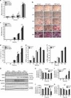
          Although denervated muscle atrophy is common, the underlying molecular mechanism remains unelucidated. We have previously found that oxidative stress and inflammatory response may be early events that trigger denervated muscle atrophy. Isoquercitrin is a biologically active flavonoid with antioxidative and anti-inflammatory properties. The present study investigated the effect of isoquercitrin on denervated soleus muscle atrophy and its possible molecular mechanisms. We found that isoquercitrin was effective in alleviating soleus muscle mass loss following denervation in a dose-dependent manner. Isoquercitrin demonstrated the optimal protective effect at 20 mg/kg/d, which was the dose used in subsequent experiments. To further explore the protective effect of isoquercitrin on denervated soleus muscle atrophy, we analyzed muscle proteolysis via the ubiquitin-proteasome pathway, mitophagy, and muscle fiber type conversion. Isoquercitrin significantly inhibited the denervation-induced overexpression of two muscle-specific ubiquitin ligases—muscle RING finger 1 (MuRF1) and muscle atrophy F-box (MAFbx), and reduced the degradation of myosin heavy chains (MyHCs) in the target muscle. Following isoquercitrin treatment, mitochondrial vacuolation and autophagy were inhibited, as evidenced by reduced level of autophagy-related proteins (ATG7, BNIP3, LC3B, and PINK1); slow-to-fast fiber type conversion in the target muscle was delayed via triggering expression of peroxisome proliferator-activated receptor γ coactivator 1α (PGC-1α); and the production of reactive oxygen species (ROS) in the target muscle was reduced, which might be associated with the upregulation of antioxidant factors (SOD1, SOD2, NRF2, NQO1, and HO1) and the downregulation of ROS production-related factors (Nox2, Nox4, and DUOX1). Furthermore, isoquercitrin treatment reduced the levels of inflammatory factors—interleukin (IL)-1β, IL-6, and tumor necrosis factor-α (TNF-α)—in the target muscle and inactivated the JAK/STAT3 signaling pathway. Overall, isoquercitrin may alleviate soleus muscle atrophy and mitophagy and reverse the slow-to-fast fiber type conversion following denervation via inhibition of oxidative stress and inflammatory response. Our study findings enrich the knowledge regarding the molecular regulatory mechanisms of denervated muscle atrophy and provide a scientific basis for isoquercitrin as a protective drug for the prevention and treatment of denervated muscle atrophy.

          Related collections

          Most cited references65

          • Record: found
          • Abstract: found
          • Article: not found

          Denervation-induced skeletal muscle atrophy is associated with increased mitochondrial ROS production.

          Reactive oxygen species (ROS), especially mitochondrial ROS, are postulated to play a significant role in muscle atrophy. We report a dramatic increase in mitochondrial ROS generation in three conditions associated with muscle atrophy: in aging, in mice lacking CuZn-SOD (Sod1(-/-)), and in the neurodegenerative disease, amyotrophic lateral sclerosis (ALS). ROS generation in muscle mitochondria is nearly threefold higher in 28- to 32-mo-old than in 10-mo-old mice and is associated with a 30% loss in gastrocnemius mass. In Sod1(-/-) mice, muscle mitochondrial ROS production is increased >100% in 20-mo compared with 5-mo-old mice along with a >50% loss in muscle mass. ALS G93A mutant mice show a 75% loss of muscle mass during disease progression and up to 12-fold higher muscle mitochondrial ROS generation. In a second ALS mutant model, H46RH48Q mice, ROS production is approximately fourfold higher than in control mice and is associated with a less dramatic loss (30%) in muscle mass. Thus ROS production is strongly correlated with the extent of muscle atrophy in these models. Because each of the models of muscle atrophy studied are associated to some degree with a loss of innervation, we were interested in determining whether denervation plays a role in ROS generation in muscle mitochondria isolated from hindlimb muscle following surgical sciatic nerve transection. Seven days post-denervation, muscle mitochondrial ROS production increased nearly 30-fold. We conclude that enhanced generation of mitochondrial ROS may be a common factor in the mechanism underlying denervation-induced atrophy.
            Bookmark
            • Record: found
            • Abstract: found
            • Article: found
            Is Open Access

            Irisin is a pro-myogenic factor that induces skeletal muscle hypertrophy and rescues denervation-induced atrophy

            Exercise induces expression of the myokine irisin, which is known to promote browning of white adipose tissue and has been shown to mediate beneficial effects following exercise. Here we show that irisin induces expression of a number of pro-myogenic and exercise response genes in myotubes. Irisin increases myogenic differentiation and myoblast fusion via activation of IL6 signaling. Injection of irisin in mice induces significant hypertrophy and enhances grip strength of uninjured muscle. Following skeletal muscle injury, irisin injection improves regeneration and induces hypertrophy. The effects of irisin on hypertrophy are due to activation of satellite cells and enhanced protein synthesis. In addition, irisin injection rescues loss of skeletal muscle mass following denervation by enhancing satellite cell activation and reducing protein degradation. These data suggest that irisin functions as a pro-myogenic factor in mice.
              Bookmark
              • Record: found
              • Abstract: found
              • Article: not found

              Trends in the design of nerve guidance channels in peripheral nerve tissue engineering.

              The current trend of peripheral nerve tissue engineering is the design of advanced nerve guidance channels (NGCs) acting as physical guidance for regeneration of nerves across lesions. NGCs should present multifunctional properties aiming to direct the sprouting of axons from the proximal nerve end, to concentrate growth factors secreted by the injured nerve ends, and to reduce the ingrowth of scar tissue into the injury site. A critical aspect in the design of NGCs is conferring them the ability to provide topographic, chemotactic and haptotactic cues that lead to functional nerve regeneration thus increasing the axon growth rate and avoiding or minimizing end-organ (e.g. muscle) atrophy. The present work reviews the recent state of the art in NGCs engineering and defines the external guide and internal fillers structural and compositional requirements that should be satisfied to improve nerve regeneration, especially in the case of large gaps (>2 cm). Techniques for NGCs fabrication were described highlighting the innovative approaches direct to enhance the regeneration of axon stumps compared to current clinical treatments. Furthermore, the possibility to apply stem cells as internal cues to the NGCs was discussed focusing on scaffold properties necessary to ensure cell survival. Finally, the optimized features for NGCs design were summarized showing as multifunctional cues are needed to produce NGCs having improved results in clinics.
                Bookmark

                Author and article information

                Contributors
                Journal
                Front Physiol
                Front Physiol
                Front. Physiol.
                Frontiers in Physiology
                Frontiers Media S.A.
                1664-042X
                12 August 2020
                2020
                : 11
                : 988
                Affiliations
                [1] 1 Key Laboratory of Neuroregeneration of Jiangsu and Ministry of Education, Jiangsu Clinical Medicine Center of Tissue Engineering and Nerve Injury Repair, Co-Innovation Center of Neuroregeneration, Nantong University , Nantong, China
                [2] 2 Department of Orthopedics, Affiliated Hospital of Nantong University , Nantong, China
                [3] 3 School of Nursing, Nantong University , Nantong, China
                Author notes

                Edited by: Capucine Trollet, Sorbonne Université, France

                Reviewed by: Bumsoo Ahn, Oklahoma Medical Research Foundation, United States; Marina Bouche, Sapienza University of Rome, Italy

                *Correspondence: Fei Ding, dingfei@ 123456ntu.edu.cn Hualin Sun, sunhl@ 123456ntu.edu.cn

                These authors have contributed equally to this work

                This article was submitted to Striated Muscle Physiology, a section of the journal Frontiers in Physiology

                Article
                10.3389/fphys.2020.00988
                7435639
                32903465
                eb8b68f7-e089-482b-8ec6-67aec8190d86
                Copyright © 2020 Shen, Zhang, Huang, Zhu, Qiu, Ma, Yang, Ding and Sun.

                This is an open-access article distributed under the terms of the Creative Commons Attribution License (CC BY). The use, distribution or reproduction in other forums is permitted, provided the original author(s) and the copyright owner(s) are credited and that the original publication in this journal is cited, in accordance with accepted academic practice. No use, distribution or reproduction is permitted which does not comply with these terms.

                History
                : 17 April 2020
                : 20 July 2020
                Page count
                Figures: 7, Tables: 0, Equations: 0, References: 75, Pages: 13, Words: 8968
                Funding
                Funded by: National Natural Science Foundation of China 10.13039/501100001809
                Award ID: 81671230
                Award ID: 81871554
                Award ID: 81901933
                Funded by: National Key Research and Development Program of China 10.13039/501100012166
                Award ID: 2017YFA0104703
                Funded by: 973 Program 10.13039/501100012166
                Award ID: 2014CB542203
                Funded by: Priority Academic Program Development of Jiangsu Higher Education Institutions (PAPD) 10.13039/501100012246
                Funded by: Nantong Science and Technology Innovation Program
                Award ID: JC2019028
                Categories
                Physiology
                Original Research

                Anatomy & Physiology
                denervated muscle atrophy,isoquercitrin,oxidative stress,inflammation,proteolysis,mitophagy

                Comments

                Comment on this article