1
views
0
recommends
+1 Recommend
0 collections
    0
    shares
      • Record: found
      • Abstract: found
      • Article: found
      Is Open Access

      Src and ROCK Kinases Differentially Regulate Mineralization of Human Osteosarcoma Saos-2 Cells

      research-article

      Read this article at

      Bookmark
          There is no author summary for this article yet. Authors can add summaries to their articles on ScienceOpen to make them more accessible to a non-specialist audience.

          Abstract

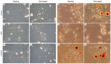
          Osteoblasts initiate bone mineralization by releasing matrix vesicles (MVs) into the extracellular matrix (ECM). MVs promote the nucleation process of apatite formation from Ca 2+ and P i in their lumen and bud from the microvilli of osteoblasts during bone development. Tissue non-specific alkaline phosphatase (TNAP) as well as annexins (among them, AnxA6) are abundant proteins in MVs that are engaged in mineralization. In addition, sarcoma proto-oncogene tyrosine-protein (Src) kinase and Rho-associated coiled-coil (ROCK) kinases, which are involved in vesicular transport, may also regulate the mineralization process. Upon stimulation in osteogenic medium containing 50 μg/mL of ascorbic acid (AA) and 7.5 mM of β-glycerophosphate (β-GP), human osteosarcoma Saos-2 cells initiated mineralization, as evidenced by Alizarin Red-S (AR-S) staining, TNAP activity, and the partial translocation of AnxA6 from cytoplasm to the plasma membrane. The addition of 4-amino-5-(4-chlorophenyl)-7-(t-butyl)pyrazolo [3,4-d] pyrimidine (PP2), which is an inhibitor of Src kinase, significantly inhibited the mineralization process when evaluated by the above criteria. In contrast, the addition of (R)-(+)-trans-4-(1-aminoethyl)-N-(4-pyridyl) cyclohexane carboxamide hydrochloride (Y-27632), which is an inhibitor of ROCK kinase, did not affect significantly the mineralization induced in stimulated Saos-2 cells as denoted by AR-S and TNAP activity. In conclusion, mineralization by human osteosarcoma Saos-2 cells seems to be differently regulated by Src and ROCK kinases.

          Related collections

          Most cited references32

          • Record: found
          • Abstract: found
          • Article: not found

          Rapid flow cytofluorometric analysis of mammalian cell cycle by propidium iodide staining

          K Krishan (1975)
          A rapid method for the flow microfluorometric determination of the DNA content per cell is described. Incubation of cells in a hypotonic solution of propidium iodide results in disruption of the cell membrane and rapid staining of nuclear chromatin. DNA distribution histograms generated from cells stained by this method are identical to those generated after fixation and RNase digestion. In contrast to some earlier described methods, the present technique is rapid (5 min of processing), requires a minimal amount of material, and avoids formation of cell clumps.
            Bookmark
            • Record: found
            • Abstract: found
            • Article: not found

            Rapidly forming apatitic mineral in an osteoblastic cell line (UMR 106-01 BSP).

            This study evaluated a rapid biomineralization phenomenon exhibited by an osteoblastic cell line, UMR 106-01 BSP, when treated with either organic phosphates [beta-glycerophosphate (beta-GP), Ser-P, or Thr-P], inorganic phosphate (P(i)), or calcium. In a dose-dependent manner, these agents (2-10 mM) stimulated confluent cultures to deposit mineral in the cell layer (ED50 of approximately 4.6 mM for beta-GP (30 +/- 2 nmol Ca2+/microgram DNA) and approximately 3.8 mM (29 +/- 2 nmol Ca2+/microgram DNA) for P(i)) with a plateau in mineral formation by 20 h (ET50 approximately 12-15 h). beta-GP or P(i) treatment yielded mineral crystals having an x-ray diffraction pattern similar to normal human bone. Alizarin red-S histology demonstrated calcium mineral deposition in the extracellular matrix and what appeared to be intracellular paranuclear staining. Electron microscopy revealed small, needle-like crystals associated with fibrillar, extracellular matrix deposits and intracellular spherical structures. Mineral formation was inhibited by levamisole (ED50 approximately 250 microM), pyrophosphate (ED50 approximately 1-10 microM), actinomycin C1 (500 ng/ml), cycloheximide (50 micrograms/ml), or brefeldin A (1 microgram/ml). These results indicate that UMR 106-01 BSP cells form a bio-apatitic mineralized matrix upon addition of supplemental phosphate. This process involves alkaline phosphatase activity, ongoing RNA and protein synthesis, as well as Golgi-mediated processing and secretion.
              Bookmark
              • Record: found
              • Abstract: found
              • Article: not found

              Annexin-mediated matrix vesicle calcification in vascular smooth muscle cells.

              In bone, osteoblasts and chondrocytes synthesize matrix vesicles (MVs) that interact with collagen to initiate calcification. MVs have been identified in human calcified arteries but are poorly characterized. The objective of this study is to determine the role of annexins and fetuin-A in MV formation and activity during calcification in bovine vascular smooth muscle cells (BVSMCs). BVSMCs were treated with control or calcification (high phosphorus) media, and cellular MVs were isolated by collagenase digestion and secreted MVs were isolated from cultured media by ultracentrifugation. The results showed that alkaline phosphatase (ALP) activity was significantly increased in MVs from calcified BVSMCs compared with noncalcified BVSMCs, as was annexin II and VI content and (45)Ca uptake. We also determined that MVs from calcifying BVSMCs could mineralize type I collagen but not type II collagen in the absence of cells in a dose- and time-dependent manner. Blockade of annexin calcium channel activity by K201 significantly decreased ALP activity and reduced the ability of the MVs to subsequently calcify on collagen, whether the K201 was added during or after MV formation. Furthermore, cellular MVs had significantly increased ability to calcify on collagen compared with secreted MVs, likely because of their increased ALP activity and annexin II content but low fetuin-A content. In conclusion, our results suggest that mineralization in VSMCs requires both active MVs and an interaction of the MVs with type I collagen, and both steps require annexin activity.
                Bookmark

                Author and article information

                Journal
                Int J Mol Sci
                Int J Mol Sci
                ijms
                International Journal of Molecular Sciences
                MDPI
                1422-0067
                12 June 2019
                June 2019
                : 20
                : 12
                : 2872
                Affiliations
                [1 ]Laboratory of Biochemistry of Lipids, Nencki Institute of Experimental Biology, Polish Academy of Sciences, 3 Pasteur Str., 02-093 Warsaw, Poland; a.strzelecka-kiliszek@ 123456nencki.gov.pl (A.S.-K.); m.romiszewska@ 123456nencki.gov.pl (M.R.); l.bozycki@ 123456nencki.gov.pl (L.B.)
                [2 ]Université de Lyon, CEDEX 69622 Villeurbanne, France; saida.mebarek@ 123456univ-lyon1.fr (S.M.); rene.buchet@ 123456univ-lyon1.fr (R.B.)
                [3 ]Université Lyon 1, CEDEX 69622 Villeurbanne, France
                [4 ]NSA de Lyon, CEDEX 69621 Villeurbanne, France
                [5 ]CPE Lyon, CEDEX 69616 Villeurbanne, France
                [6 ]ICBMS CNRS UMR 5246, CEDEX 69622 Villeurbanne, France
                [7 ]Laboratory of Cellular Metabolism, Nencki Institute of Experimental Biology, Polish Academy of Sciences, 3 Pasteur Str., 02-093 Warsaw, Poland; j.bandorowicz-pikula@ 123456nencki.gov.pl
                Author notes
                [* ]Correspondence: s.pikula@ 123456nencki.gov.pl ; Tel.: +48-22-589-2347; Fax: +48-22-822-4352
                Author information
                https://orcid.org/0000-0002-7966-3856
                https://orcid.org/0000-0003-4640-3094
                Article
                ijms-20-02872
                10.3390/ijms20122872
                6628028
                31212828
                ec3e9236-e305-4e6e-a60b-7019f491170f
                © 2019 by the authors.

                Licensee MDPI, Basel, Switzerland. This article is an open access article distributed under the terms and conditions of the Creative Commons Attribution (CC BY) license ( http://creativecommons.org/licenses/by/4.0/).

                History
                : 15 April 2019
                : 10 June 2019
                Categories
                Article

                Molecular biology
                src kinase,rock,annexin a6,mineralization,matrix vesicles,saos-2 cells
                Molecular biology
                src kinase, rock, annexin a6, mineralization, matrix vesicles, saos-2 cells

                Comments

                Comment on this article

                scite_

                Similar content604

                Cited by4

                Most referenced authors329