18
views
0
recommends
+1 Recommend
1 collections
    0
    shares
      • Record: found
      • Abstract: found
      • Article: found
      Is Open Access

      Parvalbumin-positive interneurons mediate neocortical-hippocampal interactions that are necessary for memory consolidation

      research-article

      Read this article at

      Bookmark
          There is no author summary for this article yet. Authors can add summaries to their articles on ScienceOpen to make them more accessible to a non-specialist audience.

          Abstract

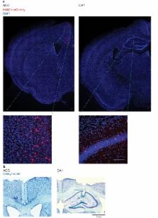
          Following learning, increased coupling between spindle oscillations in the medial prefrontal cortex (mPFC) and ripple oscillations in the hippocampus is thought to underlie memory consolidation. However, whether learning-induced increases in ripple-spindle coupling are necessary for successful memory consolidation has not been tested directly. In order to decouple ripple-spindle oscillations, here we chemogenetically inhibited parvalbumin-positive (PV +) interneurons, since their activity is important for regulating the timing of spiking activity during oscillations. We found that contextual fear conditioning increased ripple-spindle coupling in mice. However, inhibition of PV + cells in either CA1 or mPFC eliminated this learning-induced increase in ripple-spindle coupling without affecting ripple or spindle incidence. Consistent with the hypothesized importance of ripple-spindle coupling in memory consolidation, post-training inhibition of PV + cells disrupted contextual fear memory consolidation. These results indicate that successful memory consolidation requires coherent hippocampal-neocortical communication mediated by PV + cells.

          eLife digest

          Sleep contributes to the strengthening of memories. During non-dreaming sleep, neurons in regions of the brain that form and store memories – such as the hippocampus and prefrontal cortex – fire in rhythmic waves. The neurons in the hippocampus tend to fire during a wave that repeats up to 250 times per second, called sharp-wave ripples. Meanwhile, in the prefrontal cortex, the neurons tend to fire during a lower frequency wave that repeats 12 to 15 times per second, called spindles.

          During sleep and quiet wakefulness, hippocampal ripples often synchronize with prefrontal spindles; that is, both waves tend to occur at approximately the same time. Many neuroscientists think this allows the brain regions to better communicate with one another, which in turn should help the brain to strengthen memories. Consistent with this possibility, rodents that learn a new task show more synchrony between ripples and spindles afterwards. But no one had actually tested whether this increase in ripple-spindle synchrony does strengthen the rodent’s memory of the task. It was also unclear how the brain achieves such an increase.

          Xia et al. suspected that this process involved a group of inhibitory brain cells called parvalbumin-positive interneurons. These cells act like timekeepers, and help to synchronize the firing of groups of neurons. Xia et al. now show that training mice to associate an environment with a mild electric shock made it more likely that the animals would show ripple-spindle synchrony. Yet, inhibiting the activity of parvalbumin-positive interneurons in either the hippocampus or prefrontal cortex blocked this effect. It also prevented sleep from strengthening the animals’ memory of the link between the environment and the shock.

          Patients with Alzheimer’s disease have fewer parvalbumin-positive interneurons. By showing that these neurons help strengthen new memories, these findings may explain why losing them can impair memory. Restoring or replacing interneuron activity could be a promising therapeutic avenue to explore.

          Related collections

          Most cited references51

          • Record: found
          • Abstract: found
          • Article: not found

          Three groups of interneurons account for nearly 100% of neocortical GABAergic neurons.

          An understanding of the diversity of cortical GABAergic interneurons is critical to understand the function of the cerebral cortex. Recent data suggest that neurons expressing three markers, the Ca2+-binding protein parvalbumin (PV), the neuropeptide somatostatin (SST), and the ionotropic serotonin receptor 5HT3a (5HT3aR) account for nearly 100% of neocortical interneurons. Interneurons expressing each of these markers have a different embryological origin. Each group includes several types of interneurons that differ in morphological and electrophysiological properties and likely have different functions in the cortical circuit. The PV group accounts for ∼40% of GABAergic neurons and includes fast spiking basket cells and chandelier cells. The SST group, which represents ∼30% of GABAergic neurons, includes the Martinotti cells and a set of neurons that specifically target layerIV. The 5HT3aR group, which also accounts for ∼30% of the total interneuronal population, is heterogeneous and includes all of the neurons that express the neuropeptide VIP, as well as an equally numerous subgroup of neurons that do not express VIP and includes neurogliaform cells. The universal modulation of these neurons by serotonin and acetylcholine via ionotropic receptors suggests that they might be involved in shaping cortical circuits during specific brain states and behavioral contexts. Copyright © 2010 Wiley Periodicals, Inc.
            Bookmark
            • Record: found
            • Abstract: found
            • Article: not found

            Cortical interneurons that specialize in disinhibitory control

            In the mammalian cerebral cortex, the diversity of interneuronal subtypes underlies a division of labor subserving distinct modes of inhibitory control 1–7 . A unique mode of inhibitory control may be provided by inhibitory neurons that specifically suppress the firing of other inhibitory neurons. Such disinhibition could lead to the selective amplification of local processing and serve the important computational functions of gating and gain modulation 8,9 . Although several interneuron populations are known to target other interneurons to varying degrees 10–15 , little is known about interneurons specializing in disinhibition and their in vivo function. Here we show that a class of interneurons that express vasoactive intestinal polypeptide (VIP) mediates disinhibitory control in multiple areas of neocortex and is recruited by reinforcement signals. By combining optogenetic activation with single cell recordings, we examined the functional role of VIP interneurons in awake mice, and investigated the underlying circuit mechanisms in vitro in auditory and medial prefrontal cortices. We identified a basic disinhibitory circuit module in which activation of VIP interneurons transiently suppresses primarily somatostatin- and a fraction of parvalbumin-expressing inhibitory interneurons that specialize in the control of the input and output of principal cells, respectively 3,6,16,17 . During the performance of an auditory discrimination task, reinforcement signals (reward and punishment) strongly and uniformly activated VIP neurons in auditory cortex, and in turn VIP recruitment increased the gain of a functional subpopulation of principal neurons. These results reveal a specific cell-type and microcircuit underlying disinhibitory control in cortex and demonstrate that it is activated under specific behavioural conditions.
              Bookmark
              • Record: found
              • Abstract: found
              • Article: not found

              Synaptic plasticity in the anterior cingulate cortex in acute and chronic pain.

              The anterior cingulate cortex (ACC) is activated in both acute and chronic pain. In this Review, we discuss increasing evidence from rodent studies that ACC activation contributes to chronic pain states and describe several forms of synaptic plasticity that may underlie this effect. In particular, one form of long-term potentiation (LTP) in the ACC, which is triggered by the activation of NMDA receptors and expressed by an increase in AMPA-receptor function, sustains the affective component of the pain state. Another form of LTP in the ACC, which is triggered by the activation of kainate receptors and expressed by an increase in glutamate release, may contribute to pain-related anxiety.
                Bookmark

                Author and article information

                Contributors
                Role: Reviewing Editor
                Journal
                eLife
                Elife
                eLife
                eLife
                eLife Sciences Publications, Ltd
                2050-084X
                29 September 2017
                2017
                : 6
                : e27868
                Affiliations
                [1 ]deptDepartment of Physiology University of Toronto TorontoCanada
                [2 ]deptProgram in Neurosciences and Mental Health, Hospital for Sick Children University Avenue TorontoCanada
                [3 ]deptDepartment of Biological Sciences University of Toronto Scarborough TorontoCanada
                [4 ]deptDepartment of Cell and Systems Biology University of Toronto TorontoCanada
                [5 ]deptDepartment of Psychology University of Toronto TorontoCanada
                [6 ]deptInstitute of Medical Sciences University of Toronto TorontoCanada
                University of Freiburg Germany
                University of Freiburg Germany
                Author information
                http://orcid.org/0000-0001-7415-6620
                http://orcid.org/0000-0001-9662-2151
                http://orcid.org/0000-0001-5451-489X
                https://orcid.org/0000-0002-7282-7838
                http://orcid.org/0000-0002-1395-3586
                Article
                27868
                10.7554/eLife.27868
                5655147
                28960176
                ec44e0f9-6ace-4dfc-ad81-92fb4fa29032
                © 2017, Xia et al

                This article is distributed under the terms of the Creative Commons Attribution License, which permits unrestricted use and redistribution provided that the original author and source are credited.

                History
                : 17 April 2017
                : 28 September 2017
                Funding
                Funded by: FundRef http://dx.doi.org/10.13039/501100000024, Canadian Institutes of Health Research;
                Award ID: FDN143227
                Award Recipient :
                Funded by: FundRef http://dx.doi.org/10.13039/501100000024, Canadian Institutes of Health Research;
                Award ID: MOP74650
                Award Recipient :
                Funded by: FundRef http://dx.doi.org/10.13039/501100000038, Natural Sciences and Engineering Research Council of Canada;
                Award ID: RGPIN-2015-05458
                Award Recipient :
                Funded by: FundRef http://dx.doi.org/10.13039/501100000038, Natural Sciences and Engineering Research Council of Canada;
                Award ID: RGPIN-2014-04947
                Award Recipient :
                Funded by: FundRef http://dx.doi.org/10.13039/501100000038, Natural Sciences and Engineering Research Council of Canada;
                Award Recipient :
                The funders had no role in study design, data collection and interpretation, or the decision to submit the work for publication.
                Categories
                Research Article
                Neuroscience
                Custom metadata
                Electrophysiological and behavioural experiments reveal that parvalbumin interneuron-mediated increases in neocortical-hippocampal interactions are necessary for successful memory consolidation.

                Life sciences
                memory,consolidation,hippocampus,anterior cingulate cortex,ripple,spindle,mouse
                Life sciences
                memory, consolidation, hippocampus, anterior cingulate cortex, ripple, spindle, mouse

                Comments

                Comment on this article