5
views
0
recommends
+1 Recommend
0 collections
    0
    shares
      • Record: found
      • Abstract: found
      • Article: found
      Is Open Access

      Pénfigo vulgar con afectación exclusiva en el esófago: reporte de un caso Translated title: Pemphigus vulgaris with exclusive affectation in the esophagus: a case report

      research-article

      Read this article at

      Bookmark
          There is no author summary for this article yet. Authors can add summaries to their articles on ScienceOpen to make them more accessible to a non-specialist audience.

          Resumen

          Introducción:

          el pénfigo vulgar comprende un grupo de enfermedades heterogéneas autoinmunes ampollosas de la piel y las mucosas. La afectación esofágica en el pénfigo vulgar es rara, con una prevalencia incierta que requiere un abordaje diagnóstico y terapéutico detallado.

          Caso clínico:

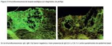
          mujer de 37 años, con antecedentes de tratamiento con inhibidores de la Cox-2 debido a hernia discal. Se envió a Gastroenterología por pérdida de peso de aprox. 5 kg en un mes. La paciente tuvo presencia de disfagia, odinofagia y dolor retroesternal con pobre tolerancia a la vía oral. Se hizo endoscopía que reportó esofagitis disecante superficial y gastropatía eritematosa de antro; el duodeno estaba en estado normal. Los hallazgos se correlacionaron con el diagnóstico de pénfigo vulgar con afectación exclusiva a esófago. En la valoración no se identificaron lesiones en piel, cavidad oral u otras mucosas. Se hizo nueva endoscopía como control y se encontró inmunofluorescencia de biopsia esofágica reactiva a IgG 2. Se dio manejo inicial con glucocorticoides, antiinflamatorios e inmunosupresores.

          Conclusiones:

          la importancia del estudio del pénfigo radica no solo en la alta morbimortalidad asociada, sino en lo raro y complejo de su detección, pues los pacientes suelen tardar varios meses en tener un diagnóstico certero y aún más en conseguir las metas terapéuticas. Es prioritaria la difusión del estudio del pénfigo entre los profesionales de la salud involucrados en su detección.

          Abstract

          Background:

          Pemphigus vulgaris comprises a group of heterogeneous blistering autoimmune diseases of the skin and mucosa. Esophageal involvement within pemphigus vulgaris is rare with an uncertain prevalence that requires a detailed diagnostic and a therapeutic approach.

          Clinical case:

          37-year-old female, with a history of treatment with Cox-2 inhibitors due to herniated disc. She is sent to the Gastroenterology Service for weight loss of approximately 5 kilos in a month, with the presence of dysphagia, odynophagia and retrosternal pain with poor tolerance to the oral route. Endoscopy was performed, which reported esophagitis dissecans superficialis (EDS), erythematous gastropathy of the antrum and normal duodenum. Findings were correlated with the diagnosis of pemphigus vulgaris with exclusive involvement of the esophagus. The evaluation did not identify lesions on the skin, oral cavity or other mucous membranes. A new endoscopy was performed as a control and it was found immunofluorescence of the esophageal biopsy reactive to IgG 2. Initial management was given with glucocorticoids, anti-inflammatories and immunosuppressants.

          Conclusions:

          The importance of the study of pemphigus lies not only in the high associated morbidity and mortality, but also in its intrinsic rarity and the complexity of its detection, given that patients usually take several months to have an accurate diagnosis and even more time to achieve therapeutic goals. It is a priority the dissemination of the study of pemphigus among health professionals involved in its detection.

          Related collections

          Most cited references22

          • Record: found
          • Abstract: found
          • Article: not found

          Diagnosis and Management of Pemphigus: recommendations by an International Panel of Experts

          Several European countries recently developed international diagnostic and management guidelines for pemphigus, which have been instrumental in the standardization of pemphigus management. We now present results from a subsequent Delphi consensus to broaden the generalizability of the recommendations. A preliminary survey, based on the European Dermatology Forum and the European Academy of Dermatology and Venereology guidelines, was sent to a panel of international experts to determine the level of consensus. The results were discussed at the International Bullous Diseases Consensus Group in March 2016 during the annual American Academy of Dermatology conference. Following the meeting, a second survey was sent to more experts to achieve greater international consensus. The 39 experts participated in the first round of the Delphi survey, and 54 experts from 21 countries completed the second round. The number of statements in the survey was reduced from 175 topics in Delphi I to 24 topics in Delphi II on the basis of Delphi results and meeting discussion. Each recommendation represents the majority opinion and therefore may not reflect all possible treatment options available. We present here the recommendations resulting from this Delphi process. This international consensus includes intravenous CD20 inhibitors as a first-line therapy option for moderate-to-severe pemphigus.
            Bookmark
            • Record: found
            • Abstract: found
            • Article: not found

            Autoimmunity to desmocollin 3 in pemphigus vulgaris.

            Pemphigus vulgaris is a blistering disease associated with autoantibodies to the desmosomal adhesion protein, desmoglein 3. Genetic deficiency of desmoglein 3 in mice mimics autoimmunity to desmoglein 3 in pemphigus vulgaris, with mucosal-dominant blistering in the suprabasal layer of the epidermis. Mice with an epidermal-specific deletion of desmocollin 3, the other major desmosomal cadherin isoform expressed in the basal epidermis, develop suprabasal blisters in skin that are histologically identical to those observed in pemphigus vulgaris, suggesting that desmocollin 3 might be a target of autoantibodies in some pemphigus vulgaris patients. We now demonstrate that desmocollin 3 is an autoantigen in pemphigus vulgaris, illustrated in a patient with mucosal-dominant blistering. Six of 38 pemphigus vulgaris and one of 85 normal serum samples immunoprecipitate desmocollin 3 (P = 0.003). Incubation of patient IgG with human keratinocytes causes loss of intercellular adhesion, and adsorption with recombinant desmocollin 3 specifically prevents this pathogenic effect. Additionally, anti-desmocollin 3 sera cause loss of keratinocyte cell surface desmocollin 3, but not desmoglein 3 by immunofluorescence, indicating distinct cellular pathogenic effects in anti-desmocollin and anti-desmoglein pemphigus, despite their identical clinical presentations. These data demonstrate that desmocollin 3 is a pathogenic autoantigen in pemphigus vulgaris and suggest that pemphigus vulgaris is a histological reaction pattern that may result from autoimmunity to desmoglein 3, desmocollin 3, or both desmosomal cadherins.
              Bookmark
              • Record: found
              • Abstract: found
              • Article: not found

              Esophagitis dissecans superficialis ("sloughing esophagitis"): a clinicopathologic study of 12 cases.

              Esophagitis dissecans superficialis (EDS) is a term applied to a rare endoscopic finding characterized by sloughing of large fragments of the esophageal squamous mucosa that may be coughed up or vomited. Although EDS has been reported in association with certain medications and esophageal strictures, most cases remain unexplained and the histopathologic features of EDS are inadequately described. We undertook this study to define useful diagnostic criteria based on the examination of a series of well-characterized cases of EDS. To identify patients with EDS, we searched our endoscopy and pathology databases, reviewed the esophageal biopsy specimens from candidate cases, and correlated them with pertinent clinical information. Twelve patients (11 men and 1 woman) had endoscopic and histologic findings of EDS and 9 had the histologic features without the endoscopic correlates. Biopsies from confirmed EDS patients showed sloughing and flaking of superficial squamous epithelium with occasional bullous separation of the layers, parakeratosis, and varying degrees of acute or chronic inflammation. Fungal elements were identified in 3 patients, but were not associated with acute inflammation. None of the EDS patients were on bisphosphonate therapy or had bullous skin disorders. Follow-up endoscopy in 5 patients showed complete resolution of the esophageal abnormalities in 4 and mild esophagitis in one. In spite of its sometimes, dramatic presentation, EDS is a benign condition that resolves without lasting esophageal pathology. Although an association with medications, skin conditions, heavy smoking, and physical trauma has been reported, the pathogenesis of EDS remains unexplained.
                Bookmark

                Author and article information

                Contributors
                Journal
                Rev Med Inst Mex Seguro Soc
                Rev Med Inst Mex Seguro Soc
                Rev Med Inst Mex Seguro Soc
                Revista Médica del Instituto Mexicano del Seguro Social
                Instituto Mexicano del Seguro Social (Ciudad de México, México )
                0443-5117
                2448-5667
                Jan-Feb 2022
                Jan-Feb 2022
                : 60
                : 1
                : 67-74
                Affiliations
                [1] originalInstituto Mexicano del Seguro Social, Unidad de Medicina Familiar No. 11, Servicio de Consulta Externa. Aguascalientes, Aguascalientes, México orgnameInstituto Mexicano del Seguro Social México
                [2] originalInstituto Mexicano del Seguro Social, Unidad de Investigación en Epidemiología y en Servicios de Salud, Área Envejecimiento. Ciudad de México, México orgnameInstituto Mexicano del Seguro Social México
                [3] originalUniversidad Autónoma de Aguascalientes, Centro de Ciencias de la Salud, Departamento de Medicina. Aguascalientes, Aguascalientes, México orgnameUniversidad Autónoma de Aguascalientes México
                [4] originalUniversidad Autónoma de Aguascalientes, Centro de Ciencias de la Salud, Departamento de Medicina. Aguascalientes, Aguascalientes, México orgnameUniversidad Autónoma de Aguascalientes México
                [5] originalUniversidad Autónoma de Aguascalientes, Centro de Ciencias de la Salud, Departamento de Medicina. Aguascalientes, Aguascalientes, México orgnameUniversidad Autónoma de Aguascalientes México
                [6] originalInstituto Mexicano del Seguro Social, Unidad de Medicina Familiar No. 8, Aguascalientes, Aguascalientes, México orgnameInstituto Mexicano del Seguro Social México
                Author notes
                [*]
                Author information
                https://orcid.org/0000-0002-9442-3471
                https://orcid.org/0000-0002-4619-6024
                https://orcid.org/0000-0001-9136-0121
                https://orcid.org/0000-0002-7238-2031
                https://orcid.org/0000-0001-8761-0765
                Article
                457770302018
                10395950
                edfdc05b-b637-4775-bc3c-50f1834bb9a8
                © 2022 Revista Medica del Instituto Mexicano del Seguro Social.

                Esta obra está bajo una Licencia Creative Commons Atribución-NoComercial-SinDerivar 4.0 Internacional.

                History
                : 10 June 2021
                : 16 November 2021
                Page count
                Figures: 5, Tables: 0, Equations: 0, References: 22
                Categories
                Casos Clínicos

                pénfigo,esófago,pemphigus,esophagus
                pénfigo, esófago, pemphigus, esophagus

                Comments

                Comment on this article