11
views
0
recommends
+1 Recommend
1 collections
    0
    shares
      • Record: found
      • Abstract: found
      • Article: found
      Is Open Access

      BjussuLAAO-II induces cytotoxicity and alters DNA methylation of cell-cycle genes in monocultured/co-cultured HepG2 cells

      research-article

      Read this article at

      ScienceOpenPublisherPMC
      Bookmark
          There is no author summary for this article yet. Authors can add summaries to their articles on ScienceOpen to make them more accessible to a non-specialist audience.

          Abstract

          Background:

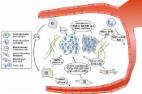
          The use of animal venoms and their toxins as material sources for biotechnological applications has received much attention from the pharmaceutical industry. L-amino acid oxidases from snake venoms (SV-LAAOs) have demonstrated innumerous biological effects and pharmacological potential against different cancer types. Hepatocellular carcinoma has increased worldwide, and the aberrant DNA methylation of liver cells is a common mechanism to promote hepatic tumorigenesis. Moreover, tumor microenvironment plays a major role in neoplastic transformation. To elucidate the molecular mechanisms responsible for the cytotoxic effects of SV-LAAO in human cancer cells, this study aimed to evaluate the cytotoxicity and the alterations in DNA methylation profiler in the promoter regions of cell-cycle genes induced by BjussuLAAO-II, an LAAO from Bothrops jaracussu venom, in human hepatocellular carcinoma (HepG2) cells in monoculture and co-culture with endothelial (HUVEC) cells.

          Methods:

          BjussuLAAO-II concentrations were 0.25, 0.50, 1.00 and 5.00 μg/mL. Cell viability was assessed by MTT assay and DNA methylation of the promoter regions of 22 cell-cycle genes by EpiTect Methyl II PCR array.

          Results:

          BjussuLAAO-II decreased the cell viability of HepG2 cells in monoculture at all concentrations tested. In co-culture, 1.00 and 5.00 μg/mL induced cytotoxicity ( p < 0.05). BjussuLAAO-II increased the methylation of CCND1 and decreased the methylation of CDKN1A in monoculture and GADD45A in both cell-culture models ( p < 0.05).

          Conclusion:

          Data showed BjussuLAAO-II induced cytotoxicity and altered DNA methylation of the promoter regions of cell-cycle genes in HepG2 cells in monoculture and co-culture models. We suggested the analysis of DNA methylation profile of GADD45A as a potential biomarker of the cell cycle effects of BjussuLAAO-II in cancer cells. The tumor microenvironment should be considered to comprise part of biotechnological strategies during the development of snake-toxin-based novel drugs.

          Related collections

          Most cited references50

          • Record: found
          • Abstract: found
          • Article: not found

          DNA methylation-based prognosis and epidrivers in hepatocellular carcinoma.

          Epigenetic deregulation has emerged as a driver in human malignancies. There is no clear understanding of the epigenetic alterations in hepatocellular carcinoma (HCC) and of the potential role of DNA methylation markers as prognostic biomarkers. Analysis of tumor tissue from 304 patients with HCC treated with surgical resection allowed us to generate a methylation-based prognostic signature using a training-validation scheme. Methylome profiling was done with the Illumina HumanMethylation450 array (Illumina, Inc., San Diego, CA), which covers 96% of known cytosine-phosphate-guanine (CpG) islands and 485,000 CpG, and transcriptome profiling was performed with Affymetrix Human Genome U219 Plate (Affymetrix, Inc., Santa Clara, CA) and miRNA Chip 2.0. Random survival forests enabled us to generate a methylation signature based on 36 methylation probes. We computed a risk score of mortality for each individual that accurately discriminated patient survival both in the training (221 patients; 47% hepatitis C-related HCC) and validation sets (n = 83; 47% alcohol-related HCC). This signature correlated with known predictors of poor outcome and retained independent prognostic capacity of survival along with multinodularity and platelet count. The subset of patients identified by this signature was enriched in the molecular subclass of proliferation with progenitor cell features. The study confirmed a high prevalence of genes known to be deregulated by aberrant methylation in HCC (e.g., Ras association [RalGDS/AF-6] domain family member 1, insulin-like growth factor 2, and adenomatous polyposis coli) and other solid tumors (e.g., NOTCH3) and describes potential candidate epidrivers (e.g., septin 9 and ephrin B2).
            Bookmark
            • Record: found
            • Abstract: found
            • Article: found
            Is Open Access

            The role of tumor microenvironment in therapeutic resistance

            Cancer cells undergo unlimited progression and survival owing to activation of oncogenes. However, support of the tumor microenvironment is essential to the formation of clinically relevant tumors. Recent evidence indicates that the tumor microenvironment is a critical regulator of immune escape, progression, and distant metastasis of cancer. Moreover, the tumor microenvironment is known to be involved in acquired resistance of tumors to various therapies. Despite significant advances in chemotherapy and radiotherapy, occurrence of therapeutic resistance leads to reduced efficacy. This review highlights myeloid cells, cancer-associated fibroblasts, and mesenchymal stem cells consisting of the tumor microenvironment, as well as the relevant signaling pathways that eventually render cancer cells to be therapeutically resistant.
              Bookmark
              • Record: found
              • Abstract: found
              • Article: found
              Is Open Access

              Genetic and Epigenetic Signatures in Human Hepatocellular Carcinoma: A Systematic Review

              Hepatocellular carcinoma (HCC) is the third most common cause of cancer deaths worldwide, and the incidence of this fatal disease is still on rise. The majority of HCCs emerge in the background of a chronic liver disease, such as chronic hepatitis and liver cirrhosis. The current understanding is that majority of HCCs evolve as a consequence of chronic inflammation and due to the presence of infection with hepatitis viruses. These underlying pathogenic stimuli subsequently induce a spectrum of genetic and epigenetic alterations in several cancer-related genes, which are involved in cell-cycle regulation, cell growth and adhesion. Such widespread genomic alterations cause disruption of normal cellular signaling and finally lead to the acquisition of a malignant phenotype in HCC. In general, the type of gene alterations, such as point mutations, deletion of chromosomal regions and abnormal methylation of gene promoters differ according to the individual targeted gene. In HCC, incidence of genetic alterations is relatively rare and is limited to a subset of few cancer-specific genes, such as the tumor suppressor p53, RB genes and oncogenes such as the CTNNB1. In contrast, epigenetic changes that involve aberrant methylation of genes and other post-transcriptional histone modifications occur far more frequently, and some of these epigenetic alterations are now being exploited for the development of molecular diagnostic signatures for HCC. In addition, recent findings of unique microRNA expression profiles also provide an evidence for the existence of novel mechanisms for gene expression regulation in HCC. In this review article, we will review the current state of knowledge on the activation of various oncogenic pathways and the inactivation of tumor suppressor pathways in HCC that result in the disruption of cancer-related gene function. In addition, we will specifically emphasize the clinical implication of some of these genetic and epigenetic alterations in the management of hepatocarcinogenesis.
                Bookmark

                Author and article information

                Journal
                J Venom Anim Toxins Incl Trop Dis
                J Venom Anim Toxins Incl Trop Dis
                jvatitd
                The Journal of Venomous Animals and Toxins Including Tropical Diseases
                Centro de Estudos de Venenos e Animais Peçonhentos - CEVAP, Universidade Estadual Paulista - UNESP
                1678-9199
                11 March 2019
                2019
                : 25
                : e147618
                Affiliations
                [1 ]Department of Clinical Analysis, Toxicology and Food Sciences, School of Pharmaceutical Sciences of Ribeirão Preto, University of São Paulo - USP, Ribeirão Preto, SP, Brazil.
                [2 ]Department of Genetics, Ribeirão Preto Medical School, University of São Paulo - USP, Ribeirão Preto, SP, Brazil.
                [3 ]Center for the Study of Venoms and Venomous Animals (CEVAP), São Paulo State University - UNESP, Botucatu, SP, Brazil.
                Author notes
                [* ]Correspondence: lusania@ 123456fcfrp.usp.br

                Competing interests: The authors declare that they have no competing interests.

                Authors’ contributions: ARTM was responsible for project development, designed the experimental approaches, interpreted the data, performed the experiments and drafted the manuscript. DLR and AFA participated in the designed experiment, interpreted data and drafted the manuscript. RSFJR and SSV provided the toxin and drafted the manuscript. LMGA coordinated and designed all the experiments, analyzed and interpreted the data, and was a major contributor in writing the manuscript. All authors read and approved the final manuscript.

                Author information
                http://orcid.org/0000-0002-3079-4388
                Article
                00305
                10.1590/1678-9199-JVATITD-1476-18
                6527400
                eeea44b9-3592-459e-82d4-18c381f6b1ae

                This article is distributed under the terms of the Creative Commons Attribution 4.0 International License ( http://creativecommons.org/licenses/by/4.0/ ), which permits unrestricted use, distribution, and reproduction in any medium, provided you give appropriate credit to the original author(s) and the source, provide a link to the Creative Commons license, and indicate if changes were made. The Creative Commons Public Domain Dedication waiver ( http://creativecommons.org/publicdomain/zero/1.0/ ) applies to the data made available in this article, unless otherwise stated.

                History
                : 29 June 2018
                : 22 October 2018
                Page count
                Figures: 3, Tables: 0, Equations: 0, References: 52, Pages: 0
                Categories
                Research

                snake venom,epigenetics,gadd45a,ccnd1,cdkn1a
                snake venom, epigenetics, gadd45a, ccnd1, cdkn1a

                Comments

                Comment on this article