23
views
0
recommends
+1 Recommend
0 collections
    0
    shares
      • Record: found
      • Abstract: found
      • Article: found
      Is Open Access

      Hyposecretion of the Adrenal Androgen Dehydroepiandrosterone Sulfate (DHEA-S) in the Majority of the Alopecia Areata Patients: Is it a Primitive and Pathogenic Perturbation of Hypothalamic-Pituitary-Adrenal Axis?

      letter

      Read this article at

      Bookmark
          There is no author summary for this article yet. Authors can add summaries to their articles on ScienceOpen to make them more accessible to a non-specialist audience.

          Abstract

          Sir, Basic and clinical research suggest that disturbed neuro-endocrine function may be involved in the pathogenesis and course of autoimmune diseases. Hormones, such as those of hypothalamic-pituitary-adrenal Axis (HPA), are known to operate a modulation of immune responses.[1] In this report, we looked into the basal serum levels of four hormones: prolactin, ACTH, cortisol, dehydroepiandrosterone sulfate (the stable metabolite of the active steroid DHEA) by means of RIA method, to investigate if it could be a gross HPA axis perturbation in severe cases of alopecia areata disease (involvement >25% of scalp hair) as it appears in other autoimmune illness, such as in rheumatoid arthritis, systemic lupus erythematosus, Sjogren disease[2] and Hashimoto's thyroiditis[3] the last one so often associated to alopecia areata.[4] We studied a total of 142 patients - 55 males and 87 females- average age 34 years, not in systemic steroid therapy [Table 1]; they are members of the “Associazione Mediterranea Alopecia Areata” (www.alopecia-italy.com). We confirmed the normal value of prolactin level[5] and did not find significant imbalance of basal level of ACTH and cortisol, but DHEA-S in the majority of the patients was found reduced in comparison to age and sex matched controls: 69.1% of males (M) and 74.7% of females (F) are below the media of the control values – 165.80±98.69 mcg/dl (M) and 101.36±97.22 mcg /dl (F) versus 228.07±151.81 mcg/dl (M- control) and 134.49±104.64 mcg/dl (F -control) - Student's t-test P<0.00002 and Mood's median-test P<0.0000001 respectively-. 63.6% of M and 64.4% of F are in the range of deficiency values - given by mean minus s.d./3: <177, 47 mcg/dl for M 
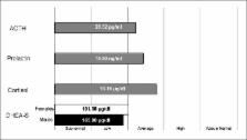
and <99,60 mcg /dl for F [Figure 1]- irrespective of their age, clinical forms and duration of the disease. At the moment, we cannot determine with certainty whether this deficit is pre-existing or subsequent to the onset of pathology, but the low DHEA-S secretion also found in the majority of the patients in the remission phase and those with recent onset of the pathology -whereas cortisol and ACTH were in the normal range- could be indicative of a primitive deficit of DHEA-S production. These results confirm the old data from Vinocurow[6] and Montagnani:[7] they found in 85% of patients a hypoadrenalism through dosing of urinary steroids, independently from the clinical form of Alopecia. Many studies have shown that DHEA/DHEA-S has significant immunomodulating activity and could be useful in restoring immune regulation in patients with chronic autoimmune diseases,[8] probably through its capacity to modulate the mechanisms of natural immunity, such as NK cells, that can control the activation of T autoreactive linphocytes, event that appears in some autoimmune diseases, including alopecia areata.[9] On the other hand, DHEA is a neurohormone with antidepressive - ansiolytic activity and low DHEA-S secretion is considered as indicative of chronic stress response,[10] whose involvement in the pathogenesis of AA is to be considered.[11] Our preliminary therapeutic data suggest the clinical usefulness in some patients of the normalitation of the defective level of DHEA-S, but it is mandatory to investigate in a consistent number of cases affected from severe chronic/relapsing AA if the administration of DHEA could be a new additive relatively safe and inexpensive resource for the stabilization of this desperating disease, as it is suggested for other autoimmune pathologies.[8] Table 1 Case study Figure 1 DHEA-S mean values in sample (normal mean and median: males=228.07±151.81 mcg/dl; females=134.49±104.64 mcg/dl): Sample mean: males=165.80±98.69 mcg/dl (n=55, Student's t-test P<0.00002), females=101.36±97.22 mcg/dl (n=87, not normally distributed, therefore, given the median=73.8, Mood's median-test P<0.0000001).

          Related collections

          Most cited references10

          • Record: found
          • Abstract: found
          • Article: not found

          Dehydroepiandrosterone as a regulator of immune cell function.

          Dehydroepiandrosterone (DHEA) is a C19 steroid of adrenal origin. Notably, its secretion declines with age, a phenomenon referred to as the "adrenopause". For many years, the physiological significance of DHEA remained elusive. However, many studies have now shown that DHEA has significant immune modulatory function, exhibiting both immune stimulatory and anti-glucocorticoid effects. Although several of these studies are limited by the fact that they were carried out in rodents, who are incapable of adrenal DHEA production, and therefore have very low circulating levels of this steroid, evidence from the study of immune cells is now accumulating to suggest a role for DHEA in regulating human immunity. This ability to regulate immune function has raised interest in the therapeutic potential of DHEA as a treatment for the immunological abnormalities that arise in subjects with low circulating levels of this hormone. This has included attempts at reversing the impaired immune response of older individuals to vaccination and restoring immune regulation in patients with chronic autoimmune disease. This review summarises the reported effects of DHEA on immune function and discusses the therapeutic potential of this steroid in geriatric medicine and particularly in age-related disease with an immune component. 2010 Elsevier Ltd. All rights reserved.
            Bookmark
            • Record: found
            • Abstract: found
            • Article: not found

            Neural immune pathways and their connection to inflammatory diseases

            Inflammation and inflammatory responses are modulated by a bidirectional communication between the neuroendocrine and immune system. Many lines of research have established the numerous routes by which the immune system and the central nervous system (CNS) communicate. The CNS signals the immune system through hormonal pathways, including the hypothalamic–pituitary–adrenal axis and the hormones of the neuroendocrine stress response, and through neuronal pathways, including the autonomic nervous system. The hypothalamic–pituitary–gonadal axis and sex hormones also have an important immunoregulatory role. The immune system signals the CNS through immune mediators and cytokines that can cross the blood–brain barrier, or signal indirectly through the vagus nerve or second messengers. Neuroendocrine regulation of immune function is essential for survival during stress or infection and to modulate immune responses in inflammatory disease. This review discusses neuroimmune interactions and evidence for the role of such neural immune regulation of inflammation, rather than a discussion of the individual inflammatory mediators, in rheumatoid arthritis.
              Bookmark
              • Record: found
              • Abstract: found
              • Article: not found

              Low serum levels of sex steroids are associated with disease characteristics in primary Sjogren's syndrome; supplementation with dehydroepiandrosterone restores the concentrations.

              Serum levels of the sex steroid prohormones dehydroepiandrosterone (DHEA) and DHEA sulfate (DHEA-S) decline upon aging and are reduced in primary Sjogren's syndrome. Our aim was to investigate: 1) effects of 50 mg oral DHEA/day on changes in serum levels of DHEA and 12 of its metabolites; 2) relationships between steroid levels and disease characteristics; and 3) whether these parameters were influenced by DHEA. Twenty-three postmenopausal women with primary Sjogren's syndrome and subnormal levels of DHEA-S were included in a randomized, 9-month, controlled, double blind crossover study. Liquid chromatography/mass spectrometry (MS)/MS and gas chromatography/MS were used to measure the sex steroids. Anti-SS-A/Ro and/or anti-SS-B/La, salivary gland focus score, salivary flow rates, dry mouth and eye symptoms, and routine laboratory tests were assessed. Baseline erythrocyte sedimentation rate was inversely correlated with testosterone (Testo), dihydrotestosterone, and DHEA-S (rs = -0.42, -0.45, and -0.58, respectively). Dry mouth symptoms correlated with low Testo and androstenedione, whereas dry eyes correlated with low estrogens, most strongly estrone (rs = -0.63). Presence of anti-SS-A and/or anti-SS-B was independently associated with low estradiol (area under the receiver operating characteristic curve, 0.82). All metabolites increased during DHEA but not during placebo. The relative increases were less for estrogens and Testo compared to dihydrotestosterone and glucuronidated androgen metabolites. Dry mouth symptoms decreased during DHEA therapy. Disease manifestations in primary Sjogren's syndrome were associated with low sex hormone levels, dry mouth symptoms with low androgens, and dry eyes with low estrogens. Exogenous DHEA was preferentially transformed into androgens rather than into estrogens.
                Bookmark

                Author and article information

                Journal
                Int J Trichology
                IJT
                International Journal of Trichology
                Medknow Publications (India )
                0974-7753
                0974-9241
                Jan-Jun 2011
                : 3
                : 1
                : 43-44
                Affiliations
                [1]National Coordinator, Trichology Group, Associazione Italiana Dermatologi Ambulatoriali (AIDA) - Day Hospital S.Camillo, Monopoli (Bari), Italy
                [1 ]Department of Statistics “C. Cecchi”, University of Bari, via C. Rosalba 53, 70124 Bari, Italy
                Author notes
                Address for correspondence: Dr. Roberto d’Ovidio, via F. Campione, 2 Bari, Italy E-mail: r.dovidio@ 123456aida.it
                Article
                IJT-3-43
                10.4103/0974-7753.82130
                3129128
                21769240
                ef059007-66f8-488a-948c-7501340f287f
                Copyright: © International Journal of Trichology

                This is an open-access article distributed under the terms of the Creative Commons Attribution-Noncommercial-Share Alike 3.0 Unported, which permits unrestricted use, distribution, and reproduction in any medium, provided the original work is properly cited.

                History
                Categories
                Letters to Editor

                Dermatology
                Dermatology

                Comments

                Comment on this article