8
views
0
recommends
+1 Recommend
0 collections
    0
    shares
      • Record: found
      • Abstract: found
      • Article: found
      Is Open Access

      Regulation of the angiotensin II-p22phox-reactive oxygen species signaling pathway, apoptosis and 8-oxoguanine-DNA glycosylase 1 retrieval in hyperoxia-induced lung injury and fibrosis in rats

      research-article

      Read this article at

      Bookmark
          There is no author summary for this article yet. Authors can add summaries to their articles on ScienceOpen to make them more accessible to a non-specialist audience.

          Abstract

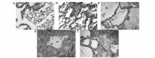
          The present study was designed to explore the impact of hyperoxia on lung injury and fibrosis via the angiotensin II (AngII)-p22phox-reactive oxygen species (ROS) signaling pathway, apoptosis and 8-oxoguanine-DNA glycosylase 1 (OGG1) repair enzyme. Newborn Sprague-Dawley rats were randomly divided in the newborn air group, newborn hyperoxia group and newborn intervention group, the latter of which was administered the chymotrypsin inhibitor, 2-(5-formylamino-6-oxo-2-phenyl-1, 6-dihydropyrimidine-1-yl)-N-[4-dioxo-1-phenyl-7-(2-pyridyloxy)] 2-heptyl-acetamide (NK3201). A group of adult rats also received hyperoxic treatment. Histomorphological changes in lung tissues were dynamically observed. AngII, ROS, angiotensin type 1 receptor ( AT 1 R) and p22phox messenger RNA (mRNA) levels, and OGG1 and peroxisome proliferator-activated receptor-γ (PPARγ) protein levels in the lung tissues were detected at various times after hyperoxia. Hyperoxia led to traumatic changes in the lungs of newborn rats that resulted in decreased viability, increased mortality, morphological changes and the apoptosis of alveolar type II epithelial cells (AT-II), as well as increased expression levels of AngII, AT 1 R and p22phox, which would ultimately lead to secondary diseases. NK3201 significantly inhibited the hyperoxia-induced increased expression of AngII, AT 1 R and p22phox and further promoted OGG1 and PPARγ protein expression, thus reducing the intrapulmonary ROS level, the apoptotic index and caspase-3 levels. However, the adult hyperoxia group only exhibited tachypnea and reduced viability. This study suggested that the AngII-p22phox-ROS signaling pathway, PPARγ and OGG1 together contributed to the hyperoxia-induced lung injury and that NK3201 was able to reverse the effects of hyperoxia.

          Related collections

          Most cited references28

          • Record: found
          • Abstract: found
          • Article: not found

          Angiotensin II-induced production of mitochondrial reactive oxygen species: potential mechanisms and relevance for cardiovascular disease.

          The role of reactive oxygen species (ROS) in angiotensin II (AngII) induced endothelial dysfunction, cardiovascular and renal remodeling, inflammation, and fibrosis has been well documented. The molecular mechanisms of AngII pathophysiological activity involve the stimulation of NADPH oxidases, which produce superoxide and hydrogen peroxide. AngII also increases the production of mitochondrial ROS, while the inhibition of AngII improves mitochondrial function; however, the specific molecular mechanisms of the stimulation of mitochondrial ROS is not clear. Interestingly, the overexpression of mitochondrial thioredoxin 2 or mitochondrial superoxide dismutase attenuates AngII-induced hypertension, which demonstrates the importance of mitochondrial ROS in AngII-mediated cardiovascular diseases. Although mitochondrial ROS plays an important role in normal physiological cell signaling, AngII, high glucose, high fat, or hypoxia may cause the overproduction of mitochondrial ROS, leading to the feed-forward redox stimulation of NADPH oxidases. This vicious cycle may contribute to the development of pathological conditions and facilitate organ damage in hypertension, atherosclerosis, and diabetes. The development of antioxidant strategies specifically targeting mitochondria could be therapeutically beneficial in these disease conditions.
            Bookmark
            • Record: found
            • Abstract: found
            • Article: not found

            Local renin-angiotensin II systems, angiotensin-converting enzyme and its homologue ACE2: their potential role in the pathogenesis of chronic obstructive pulmonary diseases, pulmonary hypertension and acute respiratory distress syndrome.

            Renin-angiotensin II-aldosterone axis has long been known as a regulator of blood pressure and fluid homeostasis. Yet, local renin-angiotensin II systems have been discovered and novel actions of angiotensin II (AngII) have emerged among which its ability to act as a immunomodulator and profibrotic molecule. The enzyme responsible for its synthesis, Angiotensin-converting-enzyme (ACE), is present in high concentrations in lung tissue. In the present paper, we review data from studies of the past decade that implicate AngII and functional polymorphisms of the ACE gene that increase ACE activity with increased susceptibility for asthma and chronic obstructive pulmonary disease (COPD) and for pulmonary hypertension. Moreover, drugs that inhibit the synthesis of AngII (ACE inhibitors) or that antagonize its actions on its receptors (Angiotensin II receptor blockers -ARBs) have been shown to provide beneficial effects. Another recent discovery reviewed is the presence of a homologue of ACE, ACE2, which cleaves a single amino acid from AngII and forms a heptapeptide with vasodilatory actions, Ang 1-7. The balance between ACE and ACE2 is crucial for controlling AngII levels. ACE and ACE2 also appear to modify the severity of Acute Respiratory Distress Syndrome (ARDS), with ACE2 playing a protective role. Finally, mention is made to the recent discovery of ACE2 as a receptor for the SARS Corona Virus.
              Bookmark
              • Record: found
              • Abstract: found
              • Article: not found

              Angiotensin-TGF-beta 1 crosstalk in human idiopathic pulmonary fibrosis: autocrine mechanisms in myofibroblasts and macrophages.

              Angiotensin II (ANGII) has been identified as a proapoptotic and profibrotic factor in experimental lung fibrosis models, and patients with the ID/DD polymorphism of ANG converting enzyme (ACE), which confers higher levels of ACE, are predisposed to lung fibrosis (Hum. Pathol. 32:521-528, 2001). Previous work from this laboratory has shown that human lung myofibroblasts isolated from patients with Idiopathic Pulmonary Fibrosis (IPF) synthesize the ANGII precursor angiotensinogen (AGT) constitutively. In attempts to understand the mechanisms and consequences of constitutive AGT synthesis by myofibroblasts, we studied myofibroblast-rich primary cultures of lung fibroblasts from patients with IPF (HIPF isolates), primary fibroblasts from normal human lung (NLFs), the IMR90 and WI38 human lung fibroblasts cell lines, and paraffin sections of lung biopsies from patients with IPF. Compared to the normal NLF isolates, HIPF primary fibroblast isolates constitutively synthesized more AGT and TGF-beta1 mRNA, and released more AGT protein, ANGII and active TGF-beta1 protein into serum-free conditioned media (both p<0.01). Incubation of HIPF fibrotic isolates with the ANGII receptor antagonist saralasin reduced both TGF-beta1 mRNA and active protein, suggesting that the constitutive expression of AGT drives the higher expression of TGF-beta1 by the HIPF cells. Consistent with this premise, treatment of either the primary NLFs or the WI38 cell line with 10(-7) M ANGII increased both TGF-beta1 mRNA and soluble active TGF-beta1 protein. Moreover, induction of the myofibroblast transition in the IMR90 cell line with 2 ng/ml TGF-beta1 increased steady state AGT mRNA levels by realtime PCR (8-fold, p<0.01) and induced expression of an AGT promoter-luciferase reporter construct by over 10-fold (p<0.001). Antisense oligonucleotides against TGF-beta1 mRNA or TGF-beta neutralizing antibodies, when applied to the fibrotic HIPF cells in serum-free medium, significantly reduced AGT expression. In lung sections from IPF patient biopsies, immunoreactive AGT/ANGI proteins were detected in myofibroblasts, epithelial cells and presumptive alveolar macrophages. Together, these data support the existence of an angiotensin/TGF-beta1 "autocrine loop" in human lung myofibroblasts and also suggest ANG peptide expression by epithelia and macrophages in the IPF lung. These findings may explain the ability of ACE inhibitors and ANG receptor antagonists to block experimental lung fibrosis in animals, and support the need for evaluation of these agents for potential treatment of human IPF. This manuscript discusses the data described above and their implications regarding IPF pathogenesis.
                Bookmark

                Author and article information

                Journal
                Exp Ther Med
                Exp Ther Med
                ETM
                Experimental and Therapeutic Medicine
                D.A. Spandidos
                1792-0981
                1792-1015
                June 2017
                05 May 2017
                05 May 2017
                : 13
                : 6
                : 3397-3407
                Affiliations
                [1 ]Department of Pediatrics, Southwest Hospital of The Third Military Medical University, Chongqing 400038, P.R. China
                [2 ]Department of Oncology, The First Affiliated Hospital of Chongqing Medical University, Chongqing 400016, P.R. China
                [3 ]Department of Pharmacy, Children's Hospital of Chongqing Medical University, Chongqing 400014, P.R. China
                [4 ]Pediatric Intensive Care Unit, Children's Hospital of Chongqing Medical University, Chongqing 400014, P.R. China
                Author notes
                Correspondence to: Dr Yuxi Zhu, Department of Oncology, The First Affiliated Hospital of Chongqing Medical University, 1 Youyi Road, Yuzhong, Chongqing 400016, P.R. China, E-mail: yuxizhucn@ 123456126.com
                Professor Feng Xu, Pediatric Intensive Care Unit, Children's Hospital of Chongqing Medical University, 136 Zhongshan Er Road, Yuzhong, Chongqing 400014, P.R. China, E-mail: xuf2011@ 123456yeah.net
                Article
                ETM-0-0-4429
                10.3892/etm.2017.4429
                5450571
                f05a28cf-9923-458b-8701-84f366d87ed9
                Copyright: © Wang et al.

                This is an open access article distributed under the terms of the Creative Commons Attribution-NonCommercial-NoDerivs License, which permits use and distribution in any medium, provided the original work is properly cited, the use is non-commercial and no modifications or adaptations are made.

                History
                : 15 August 2015
                : 13 January 2017
                Categories
                Articles

                Medicine
                oxidative stress,angiotensin ii,nk3201,human 8-oxoguanine-dna glycosylase 1,peroxisome proliferator-activated receptor-γ

                Comments

                Comment on this article