16
views
0
recommends
+1 Recommend
0 collections
    0
    shares
      • Record: found
      • Abstract: found
      • Article: found
      Is Open Access

      The Prognostic Value of Residual Volume/Total Lung Capacity in Patients with Chronic Obstructive Pulmonary Disease

      research-article

      Read this article at

      Bookmark
          There is no author summary for this article yet. Authors can add summaries to their articles on ScienceOpen to make them more accessible to a non-specialist audience.

          Abstract

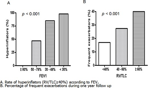
          The prognostic role of resting pulmonary hyperinflation as measured by residual volume (RV)/total lung capacity (TLC) in chronic obstructive pulmonary disease (COPD) remains poorly understood. Therefore, this study aimed to identify the factors related to resting pulmonary hyperinflation in COPD and to determine whether resting pulmonary hyperinflation is a prognostic factor in COPD. In total, 353 patients with COPD in the Korean Obstructive Lung Disease cohort recruited from 16 hospitals were enrolled. Resting pulmonary hyperinflation was defined as RV/TLC ≥ 40%. Multivariate logistic regression analysis demonstrated that older age ( P = 0.001), lower forced expiratory volume in 1 second (FEV 1) ( P < 0.001), higher St. George Respiratory Questionnaire (SGRQ) score ( P = 0.019), and higher emphysema index ( P = 0.010) were associated independently with resting hyperinflation. Multivariate Cox regression model that included age, gender, dyspnea scale, SGRQ, RV/TLC, and 6-min walking distance revealed that an older age (HR = 1.07, P = 0.027), a higher RV/TLC (HR = 1.04, P = 0.025), and a shorter 6-min walking distance (HR = 0.99, P < 0.001) were independent predictors of all-cause mortality. Our data showed that older age, higher emphysema index, higher SGRQ score, and lower FEV 1 were associated independently with resting pulmonary hyperinflation in COPD. RV/TLC is an independent risk factor for all-cause mortality in COPD.

          Graphical Abstract

          Related collections

          Most cited references16

          • Record: found
          • Abstract: found
          • Article: not found

          Susceptibility to exacerbation in chronic obstructive pulmonary disease.

          Although we know that exacerbations are key events in chronic obstructive pulmonary disease (COPD), our understanding of their frequency, determinants, and effects is incomplete. In a large observational cohort, we tested the hypothesis that there is a frequent-exacerbation phenotype of COPD that is independent of disease severity. We analyzed the frequency and associations of exacerbation in 2138 patients enrolled in the Evaluation of COPD Longitudinally to Identify Predictive Surrogate Endpoints (ECLIPSE) study. Exacerbations were defined as events that led a care provider to prescribe antibiotics or corticosteroids (or both) or that led to hospitalization (severe exacerbations). Exacerbation frequency was observed over a period of 3 years. Exacerbations became more frequent (and more severe) as the severity of COPD increased; exacerbation rates in the first year of follow-up were 0.85 per person for patients with stage 2 COPD (with stage defined in accordance with Global Initiative for Chronic Obstructive Lung Disease [GOLD] stages), 1.34 for patients with stage 3, and 2.00 for patients with stage 4. Overall, 22% of patients with stage 2 disease, 33% with stage 3, and 47% with stage 4 had frequent exacerbations (two or more in the first year of follow-up). The single best predictor of exacerbations, across all GOLD stages, was a history of exacerbations. The frequent-exacerbation phenotype appeared to be relatively stable over a period of 3 years and could be predicted on the basis of the patient's recall of previous treated events. In addition to its association with more severe disease and prior exacerbations, the phenotype was independently associated with a history of gastroesophageal reflux or heartburn, poorer quality of life, and elevated white-cell count. Although exacerbations become more frequent and more severe as COPD progresses, the rate at which they occur appears to reflect an independent susceptibility phenotype. This has implications for the targeting of exacerbation-prevention strategies across the spectrum of disease severity. (Funded by GlaxoSmithKline; ClinicalTrials.gov number, NCT00292552.)
            Bookmark
            • Record: found
            • Abstract: found
            • Article: not found

            Computed tomographic measurements of airway dimensions and emphysema in smokers. Correlation with lung function.

            Chronic obstructive pulmonary disease (COPD) is characterized by the presence of airflow obstruction caused by emphysema or airway narrowing, or both. Low attenuation areas (LAA) on computed tomography (CT) have been shown to represent macroscopic or microscopic emphysema, or both. However CT has not been used to quantify the airway abnormalities in smokers with or without airflow obstruction. In this study, we used CT to evaluate both emphysema and airway wall thickening in 114 smokers. The CT measurements revealed that a decreased FEV(1) (%predicted) is associated with an increase of airway wall area and an increase of emphysema. Although both airway wall thickening and emphysema (LAA) correlated with measurements of lung function, stepwise multiple regression analysis showed that the combination of airway and emphysema measurements improved the estimate of pulmonary function test abnormalities. We conclude that both CT measurements of airway dimensions and emphysema are useful and complementary in the evaluation of the lung of smokers.
              Bookmark
              • Record: found
              • Abstract: found
              • Article: not found

              Comparison of computed density and macroscopic morphometry in pulmonary emphysema.

              High-resolution computed tomography (HRCT) scans were obtained at 1 cm intervals in 63 subjects referred for surgical resection of a cancer or for transplantation to find out whether the relative area of lung occupied by attenuation values lower than a threshold would be a measurement of macroscopic emphysema. Using a semiautomatic procedure, the relative areas occupied by attenuation values lower than eight thresholds ranging from -900 to -970 HU were calculated on the set of scans obtained through the lobe or the lung to be resected. The extent of emphysema was quantified by a computer-assisted method on horizontal paper-mounted lung sections obtained every 1 to 2 cm. The only level for which no statistically significant difference was found between the HRCT and the morphometric data was -950 HU. To determine the number of scans sufficient for an accurate quantification, we recalculated the relative area occupied by attenuation values lower than -950 HU on progressively fewer numbers of scans and investigated the departure from the results obtained with 1 cm intervals. Because of wide variations in this departure from patient to patient, a standard cannot be recommended as the optimal distance between scans.
                Bookmark

                Author and article information

                Journal
                J Korean Med Sci
                J. Korean Med. Sci
                JKMS
                Journal of Korean Medical Science
                The Korean Academy of Medical Sciences
                1011-8934
                1598-6357
                October 2015
                12 September 2015
                : 30
                : 10
                : 1459-1465
                Affiliations
                [1 ]Division of Pulmonology, Allergy & Critical Care Medicine, Department of Internal Medicine, Hallym University Medical Center, Seoul, Korea.
                [2 ]Department of Pulmonary and Critical Care Medicine and Clinical Research Center for Chronic Obstructive Airway Diseases, Asan Medical Center, University of Ulsan College of Medicine, Seoul, Korea.
                [3 ]Department of Pulmonary and Critical Care Medicine, Ajou University School of Medicine, Suwon, Korea.
                [4 ]Office of Biostatistics, Medical Humanities and Social Medicine, Ajou University School of Medicine, Seoul, Korea.
                [5 ]Department of Radiology, Asan Medical Center, University of Ulsan College of Medicine, Seoul, Korea.
                [6 ]Department of Internal Medicine, Konkuk University School of Medicine, Seoul, Korea.
                [7 ]Department of Internal Medicine, CHA Bundang Medical Center, CHA University, Seongnam, Korea.
                [8 ]Division of Pulmonology, Department of Internal Medicine, Hanyang University Guri Hospital, Hanyang University College of Medicine, Guri, Korea.
                [9 ]Division of Pulmonary and Critical Care Medicine, Department of Medicine, Kangbuk Samsung Hospital, Sungkyunkwan University School of Medicine, Seoul, Korea.
                [10 ]Department of Internal Medicine, Seoul National University Bundang Hospital, Seoul National University College of Medicine, Seongnam, Korea.
                [11 ]Department of Internal Medicine, Seoul St. Mary's Hospital, Catholic University of Korea, Seoul, Korea.
                [12 ]Division of Pulmonary and Critical Care Medicine, Department of Internal Medicine, College of Medicine, Chungbuk National University, Cheongju, Korea.
                Author notes
                Address for Correspondence: Joo Hun Park, MD. Department of Pulmonary and Critical Care Medicine, Ajou University School of Medicine, 164 Worldcup-ro, Yeongtong-gu, Suwon 16499, Korea. Tel: +82.31-219-5116, Fax: +82.31-219-5124, jhpamc@ 123456hanmail.net

                *Tae Rim Shin and Yeon-Mok Oh contributed equally to this work.

                Article
                10.3346/jkms.2015.30.10.1459
                4575935
                26425043
                f0831576-7b93-4070-8dad-c19084fe1aab
                © 2015 The Korean Academy of Medical Sciences.

                This is an Open Access article distributed under the terms of the Creative Commons Attribution Non-Commercial License ( http://creativecommons.org/licenses/by-nc/4.0/) which permits unrestricted non-commercial use, distribution, and reproduction in any medium, provided the original work is properly cited.

                History
                : 27 March 2015
                : 10 July 2015
                Funding
                Funded by: Ministry of Health and Welfare
                Award ID: A102065
                Award ID: HI10C2020
                Categories
                Original Article
                Respiratory Diseases

                Medicine
                copd,hyperinflation,survival,residual volume,total lung capacity
                Medicine
                copd, hyperinflation, survival, residual volume, total lung capacity

                Comments

                Comment on this article