Inviting an author to review:
Find an author and click ‘Invite to review selected article’ near their name.
Search for authorsSearch for similar articles
9
views
0
recommends
+1 Recommend
0 collections
    0
    shares
      • Record: found
      • Abstract: found
      • Article: found
      Is Open Access

      Reduced functional connectivity of somatosensory network in writer's cramp patients

      research-article

      Read this article at

      Bookmark
          There is no author summary for this article yet. Authors can add summaries to their articles on ScienceOpen to make them more accessible to a non-specialist audience.

          Abstract

          Background

          The involvement of motor cortex and sensorimotor integration in patients with writer's cramp ( WC) has been well documented. However, the exact neurophysiological profile within the somatosensory system, including primary somatosensory cortex ( SI), contralateral ( SIIc), and ipsilateral ( SIIi) secondary somatosensory areas remains less understood.

          Methods

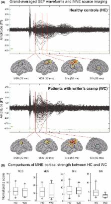
          This study investigated the neuromagnetic cortical activities of median nerve stimulation in 10 patients with WC and 10 healthy controls ( HC). To comprehensively explore all the aspects of somatosensory functioning, we analyzed our data with the minimum norm estimate ( MNE), the time‐frequency approach with evoked and induced activities, and functional connectivity between SI and SIIc ( SISIIc), SI and SIIi ( SISIIi), and SIIc and SIIi ( SIIc– SIIi) from theta to gamma oscillations.

          Results

          No significant between‐group differences were found in the MNE cortical amplitudes of SI, SIIc, and SIIi. Power strengths of evoked gamma oscillation and induced beta synchronization were also equivalent between WC and HC groups. However, we found significantly reduced theta coherence of SISIIi, alpha coherence of SISIIi and SIIc– SIIi, as well as beta coherence of SIIc– SIIi in patients with WC.

          Conclusion

          Our results suggest the involvement of somatosensory abnormalities, primarily with the form of functional connectivity, in patients with WC.

          Related collections

          Most cited references23

          • Record: found
          • Abstract: found
          • Article: not found

          Functional segregation of movement-related rhythmic activity in the human brain.

          Multiple synaptic interconnections in the human brain support concerted rhythmic activity of a large number of cortical neurons, typically close to 10 and 20 Hz. Our present neuromagnetic data provide evidence for distinct functional roles of these spectral components in the somatomotor cortex. The sites of suppression during movement and the subsequent rebound of the 20-Hz rhythm followed, along the motor cortex, the representation of fingers, toes, and mouth, as opposed to the stable origin of the 10-Hz rhythms close to the hand somatosensory cortex. The 20-Hz activity appears to be a signature of active immobilization following movement, whereas the reactive 10-Hz signals likely reflect lack of relevant sensory input from the important upper limbs.
            Bookmark
            • Record: found
            • Abstract: found
            • Article: not found

            Activation of human primary motor cortex during action observation: a neuromagnetic study.

            The monkey premotor cortex contains neurons that discharge during action execution and during observation of actions made by others. Transcranial magnetic stimulation experiments suggest that a similar observation/execution matching system also is present in humans. We recorded neuromagnetic oscillatory activity of the human precentral cortex from 10 healthy volunteers while (i) they had no task to perform, (ii) they were manipulating a small object, and (iii) they were observing another individual performing the same task. The left and right median nerves were stimulated alternately (interstimulus interval, 1.5 s) at intensities exceeding motor threshold, and the poststimulus rebound of the rolandic 15- to 25-Hz activity was quantified. In agreement with previous studies, the rebound was strongly suppressed bilaterally during object manipulation. Most interestingly, the rebound also was significantly diminished during action observation (31-46% of the suppression during object manipulation). Control experiments, in which subjects were instructed to observe stationary or moving stimuli, confirmed the specificity of the suppression effect. Because the recorded 15- to 25-Hz activity is known to originate mainly in the precentral motor cortex, we concluded that the human primary motor cortex is activated during observation as well as execution of motor tasks. These findings have implications for a better understanding of the machinery underlying action recognition in humans.
              Bookmark
              • Record: found
              • Abstract: found
              • Article: not found

              The pathophysiology of primary dystonia.

              Co-contraction and overflow of EMG activity of inappropriate muscles are typical features of all dystonic movements whether voluntary or involuntary. Voluntary movements are slow and more variable than normal, and there is particular difficulty switching between component movements of a complex task. Reduced spinal cord and brainstem inhibition is common to many reflex studies (long-latency reflexes, cranial reflexes and reciprocal inhibition). These reflex abnormalities may contribute to the difficulties in voluntary movements but cannot be causal as they can occur outside the clinically involved territory. Clinical and neurophysiological studies have emphasized the possible role of sensory feedback in the generation of dystonic movements. Abnormalities of cortical and basal ganglia function have been described in functional imaging and neurophysiological studies of patients with dystonia and in animal models of primary dystonia. Studies of cortical function have shown reduced preparatory activity in the EEG before the onset of voluntary movements, whilst magnetic brain stimulation has revealed changes in motor cortical excitability. Functional imaging of the brain in primary dystonia has suggested reduced pallidal inhibition of the thalamus with consequent overactivity of medial and prefrontal cortical areas and underactivity of the primary motor cortex during movements. These findings are supported by preliminary neuronal recordings from the globus pallidus and the thalamus at the time of stereotaxic surgery in patients with dystonia. All this evidence suggests that primary dystonia results from a functional disturbance of the basal ganglia, particularly in the striatal control of the globus pallidus (and substantia nigra pars reticulata). This causes altered thalamic control of cortical motor planning and executive areas, and abnormal regulation of brainstem and spinal cord inhibitory interneuronal mechanisms.
                Bookmark

                Author and article information

                Journal
                Brain Behav
                Brain Behav
                10.1002/(ISSN)2157-9032
                BRB3
                Brain and Behavior
                John Wiley and Sons Inc. (Hoboken )
                2162-3279
                26 January 2016
                March 2016
                : 6
                : 3 ( doiID: 10.1002/brb3.2016.6.issue-3 )
                : e00433
                Affiliations
                [ 1 ] Department of Occupational Therapy Graduate Institute of Behavioral SciencesChang Gung University TaoyuanTaiwan
                [ 2 ] Healthy Aging Research CenterChang Gung University TaoyuanTaiwan
                [ 3 ] Department of PsychiatryChang Gung Memorial Hospital TaoyuanTaiwan
                [ 4 ] Institute of PhysiologyNational Yang‐Ming University TaipeiTaiwan
                [ 5 ] Laboratory of NeurophysiologyTaipei Veterans General Hospital TaipeiTaiwan
                [ 6 ] Department of NeurologyChang Gung Memorial Hospital TaoyuanTaiwan
                [ 7 ] College of MedicineChang Gung University TaoyuanTaiwan
                [ 8 ] Institute of Brain ScienceNational Yang‐Ming University TaipeiTaiwan
                [ 9 ] Department of NeurologyTaipei Veterans General Hospital TaipeiTaiwan
                Author notes
                [*] [* ] Correspondence

                Chia‐Hsiung Cheng, Department of Occupational Therapy and Graduate Institute of Behavioral Sciences, Chang Gung University, No. 259, Wenhua 1st Rd., Taoyuan City 333, Taiwan. Tel: +886 3 2118800 #3854; Fax: +886 3 2118700; E‐mails: ch.cheng@ 123456mail.cgu.edu.tw , chiahsiung.cheng@ 123456gmail.com

                and

                Yung‐Yang Lin, Institute of Brain Science, National Yang‐Ming University, and Department of Neurology, Taipei Veterans General Hospital, No.201, Sec.2, Shipai Rd., Taipei City 112, Taiwan. Tel: +886 2 28757398; Fax: +886 2 28757579; E‐mails: yylin@ 123456vghtpe.gov.tw , g2000kev@ 123456gmail.com

                Article
                BRB3433
                10.1002/brb3.433
                4726822
                26839735
                f2d1d9ac-75b5-40e2-8355-67209d9fd8aa
                © 2016 The Authors. Brain and Behavior published by Wiley Periodicals, Inc.

                This is an open access article under the terms of the Creative Commons Attribution License, which permits use, distribution and reproduction in any medium, provided the original work is properly cited.

                History
                : 11 August 2015
                : 07 September 2015
                : 16 December 2015
                Page count
                Pages: 8
                Funding
                Funded by: Taipei Veterans General Hospital
                Award ID: V104C‐068
                Award ID: V104E9‐004
                Award ID: V103C‐062
                Award ID: V103E9‐003
                Funded by: Chang Gung Memorial Hospital
                Award ID: CMRPD1E0291
                Funded by: Chang Gung University
                Award ID: BMRPE25
                Funded by: Healthy Aging Research Center at Chang Gung University
                Award ID: EMRPD1D0291
                Award ID: CMRPD1B0331
                Award ID: EMRPD1E1711
                Funded by: National Science Council
                Award ID: NSC‐102‐2628‐B‐010‐008‐MY3
                Award ID: NSC‐101‐2314‐B‐010‐068‐MY3
                Funded by: Ministry of Science and Technology
                Award ID: MOST‐104‐2314‐B‐182‐001‐MY2
                Categories
                Original Research
                Original Research
                Custom metadata
                2.0
                brb3433
                March 2016
                Converter:WILEY_ML3GV2_TO_NLMPMC version:4.8.6 mode:remove_FC converted:12.04.2016

                Neurosciences
                coherence,dystonia,magnetoencephalography (meg),median nerve stimulation,secondary somatosensory cortex (sii)

                Comments

                Comment on this article