1
views
0
recommends
+1 Recommend
0 collections
    0
    shares
      • Record: found
      • Abstract: found
      • Article: found
      Is Open Access

      Logistic regression models of cytokines in differentiating vitreoretinal lymphoma from uveitis

      research-article

      Read this article at

      Bookmark
          There is no author summary for this article yet. Authors can add summaries to their articles on ScienceOpen to make them more accessible to a non-specialist audience.

          Abstract

          Background

          Vitreoretinal lymphoma (VRL) can commonly masquerade as chronic idiopathic uveitis due to its nonspecific clinical presentation. Thus, its early diagnosis is difficult. In this study, new logistic regression models were used to classify VRL and uveitis. Additionally, the diagnostic performance of interleukin (IL)‐10, the IL‐10/IL‐6, and the Interleukin Score for IntraOcular Lymphoma Diagnosis (ISOLD) are evaluated.

          Methods

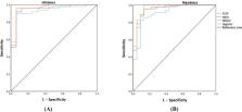
          Sixty‐nine aqueous humors (AH) (46 VRL, 23 uveitis) and 65 vitreous humors (VH) (49 VRL, 16 uveitis) were collected from a single‐center retrospective cohort. Logistic regression models were conducted based on IL‐6 and IL‐10. The cut‐off values, area under the receiver operating characteristic curve (ROC) curve (AUC), sensitivity and specificity of IL‐10, the IL‐10/IL‐6, the ISOLD, and the models were calculated from the ROC. Furthermore, Spearman's rank correlation analysis was performed to determine cytokine levels in VH and AH.

          Results

          We redefined the cut‐off values of IL‐10, the IL‐10/IL‐6, the ISOLD, and the logistic regression models. In AH, the AUC values of IL‐10, ISOLD, IL10/IL6, and the model were 0.91, 0.953, 0.952, and 0.967. In VH, they were 0.93, 0.95, 0.954, and 0.954, respectively. IL‐6 ( r = 0.7844) and IL‐10 ( r = 0.8506) in AH and VH showed a strong correlation.

          Conclusions

          IL‐6 and IL‐10 levels were introduced into new logistic regression models. The diagnostic efficacy of the models improved compared to the indicators mentioned above among Chinese patients. Additionally, the models could predict the probability of VRL more accurately. A strong correlation of cytokine levels showed the great potential of AH as prioritized auxiliary diagnostic for VRL.

          Abstract

          The ROC curve analysis ofthe IL‐10 levels, the IL‐10/IL‐6 ratio, the ISOLD and the logistic regression model indiagnosis for lymphoma.

          Related collections

          Most cited references27

          • Record: found
          • Abstract: found
          • Article: not found

          Primary vitreoretinal lymphoma: a report from an International Primary Central Nervous System Lymphoma Collaborative Group symposium.

          Primary vitreoretinal lymphoma (PVRL), also known as primary intraocular lymphoma, is a rare malignancy typically classified as a diffuse large B-cell lymphoma and most frequently develops in elderly populations. PVRL commonly masquerades as posterior uveitis and has a unique tropism for the retina and central nervous system (CNS). Over 15% of primary CNS lymphoma patients develop intraocular lymphoma, usually occurring in the retina and/or vitreous. Conversely, 65%-90% of PVRL patients develop CNS lymphoma. Consequently, PVRL is often fatal because of ultimate CNS association. Current PVRL animal models are limited and require further development. Typical clinical findings include vitreous cellular infiltration (lymphoma and inflammatory cells) and subretinal tumor infiltration as determined using dilated fundoscopy, fluorescent angiography, and optical coherent tomography. Currently, PVRL is most often diagnosed using both histology to identify lymphoma cells in the vitreous or retina and immunohistochemistry to indicate monoclonality. Additional adjuncts in diagnosing PVRL exist, including elevation of interleukin-10 levels in ocular fluids and detection of Ig(H) or T-cell receptor gene rearrangements in malignant cells. The optimal therapy for PVRL is not defined and requires the combined effort of oncologists and ophthalmologists. PVRL is sensitive to radiation therapy and exhibits high responsiveness to intravitreal methotrexate or rituximab. Although systemic chemotherapy alone can result in high response rates in patients with PVRL, there is a high relapse rate. Because of the disease rarity, international, multicenter, collaborative efforts are required to better understand the biology and pathogenesis of PVRL as well as to define both diagnostic markers and optimal therapies.
            Bookmark
            • Record: found
            • Abstract: found
            • Article: not found

            Clinical features and diagnostic significance of the intraocular fluid of 217 patients with intraocular lymphoma.

            Intraocular lymphoma is a rare disease with a poor prognosis. Early diagnosis and early treatment greatly influence the survival prognosis of this disease. This retrospective study aimed to clarify the clinical features of patients diagnosed with intraocular lymphoma, and the diagnostic significance of results from analysis of vitreous samples including cytology, cytokine measurements, and the IgH gene rearrangement test. We reviewed 217 patients with intraocular lymphoma diagnosed at 25 medical institutions in Japan. Together with clinical observation, cytological analysis, determination of the levels of cytokines, and/or detection of IgH gene rearrangements were conducted using vitreous fluid specimens. The results were studied in conjunction with clinical findings of intraocular lymphoma. Survival curves were estimated by use of the Kaplan-Meier method. The subjects comprised 85 men and 132 women, with a mean age at first ophthalmological examination of 63.4 years. The mean observation period was 41.3 months. During the observation period, 69 patients had onset of lymphoma in one eye and 148 had onset in both eyes. Intraocular lymphoma with involvement of the central nervous system (CNS) was most common, found in 60.8 % of the patients, whereas intraocular lymphoma without involvement of other organs was found in 28.1 % of patients. With respect to onset patterns, 82.5 % of patients developed primary ocular lesions whereas 16.1 % developed primary CNS lesions preceding intraocular lymphoma. Blurred vision and ataxia were the most common ocular and extra-ocular symptoms that prompted patients to seek medical examination. Vitreous opacification was the most common ocular finding. The detection rates of malignant cytology, IL-10/IL-6 ratio greater than 1.0, and IgH gene rearrangements in vitreous specimens were 44.5, 91.7, and 80.6 %, respectively, of patients tested. IL-10/IL-6 ratio greater than 1.0 had the highest overall detection rate, and was extremely high (≥90 %) in patients with or without vitreous opacification. The 5-year survival rate was 61.1 %. Cytokine analysis of vitreous biopsy had the highest detection rate for intraocular lymphoma. This supplementary diagnostic test should be performed frequently to confirm a diagnosis of intraocular lymphoma.
              Bookmark
              • Record: found
              • Abstract: found
              • Article: not found

              Primary intraocular lymphoma.

              Primary intraocular lymphoma (PIOL) is an ocular malignancy that is a subset of primary central system lymphoma (PCNSL). Approximately one-third of PIOL patients will have concurrent PCNSL at presentation, and 42-92% will develop PCNSL within a mean of 8-29 months. Although rare, the incidence has been rising in both immunocompromised and immunocompetent populations. The majority of PIOL is diffuse large B-cell lymphoma, though rare T-cell variants are described. Recently, PIOL has been classified by main site of involvement in the eye, with vitreoretinal lymphoma as the most common type of ocular lymphoma related to PCNSL. Diagnosis remains challenging for ophthalmologists and pathologists. PIOL can masquerade as noninfectious or infectious uveitis, white dot syndromes, or occasionally as other neoplasms such as metastatic cancers. Laboratory diagnosis by cytology has been much aided by the use of immunocytochemistry, flow cytometry, biochemical finding of interleukin changes (IL10:IL6 ratio > 1), and cellular microdissection with polymerase chain reaction amplification for clonality. Use of several tests improves the diagnostic yield. Approaches to treatment have centered on systemic methotrexate-based chemotherapy, often with cytarabine (Ara-C) and radiotherapy. Use of intravitreal chemotherapy with methotrexate (0.4 mg/0.1 mL) is promising in controlling ocular disease, and intravitreal rituximab (anti-CD20 monoclonal antibody) has also been tried. Despite these advances, prognosis remains poor.
                Bookmark

                Author and article information

                Contributors
                wangqingping71@163.com
                guanming88@126.com
                Journal
                J Clin Lab Anal
                J Clin Lab Anal
                10.1002/(ISSN)1098-2825
                JCLA
                Journal of Clinical Laboratory Analysis
                John Wiley and Sons Inc. (Hoboken )
                0887-8013
                1098-2825
                13 September 2022
                October 2022
                : 36
                : 10 ( doiID: 10.1002/jcla.v36.10 )
                : e24689
                Affiliations
                [ 1 ] Department of Ophthalmology, Huashan Hospital Fudan University Shanghai China
                [ 2 ] Department of Ophthalmology, Jinshan Hospital Fudan University Shanghai China
                [ 3 ] Department of Laboratory Medicine, Huashan Hospital Fudan University Shanghai China
                [ 4 ] Department of Hematology, Huashan Hospital Fudan University Shanghai China
                Author notes
                [*] [* ] Correspondence

                Qingping Wang, Department of Ophthalmology, Huashan Hospital, Fudan University, Shanghai 200040, China.

                Email: wangqingping71@ 123456163.com

                Ming Guan, Department of Laboratory Medicine, Huashan Hospital, Fudan University, Shanghai 200040, China.

                Email: guanming88@ 123456126.com

                Author information
                https://orcid.org/0000-0002-5336-7573
                Article
                JCLA24689 JCLA-22-2049.R1
                10.1002/jcla.24689
                9550960
                36098043
                f380d7b8-f99d-4229-be5b-e75638663c73
                © 2022 The Authors. Journal of Clinical Laboratory Analysis published by Wiley Periodicals LLC.

                This is an open access article under the terms of the http://creativecommons.org/licenses/by-nc-nd/4.0/ License, which permits use and distribution in any medium, provided the original work is properly cited, the use is non‐commercial and no modifications or adaptations are made.

                History
                : 18 August 2022
                : 18 July 2022
                : 25 August 2022
                Page count
                Figures: 3, Tables: 5, Pages: 7, Words: 4447
                Categories
                Research Article
                Research Articles
                Custom metadata
                2.0
                October 2022
                Converter:WILEY_ML3GV2_TO_JATSPMC version:6.2.0 mode:remove_FC converted:11.10.2022

                Clinical chemistry
                il‐6,il‐10,isold,logistic regression models,uveitis,vitreoretinal lymphoma
                Clinical chemistry
                il‐6, il‐10, isold, logistic regression models, uveitis, vitreoretinal lymphoma

                Comments

                Comment on this article