30
views
0
recommends
+1 Recommend
0 collections
    0
    shares
      • Record: found
      • Abstract: found
      • Article: not found

      High-density lipoprotein induces cyclooxygenase-2 expression and prostaglandin I-2 release in endothelial cells through sphingosine kinase-2

      research-article

      Read this article at

      ScienceOpenPublisherPMC
      Bookmark
          There is no author summary for this article yet. Authors can add summaries to their articles on ScienceOpen to make them more accessible to a non-specialist audience.

          Abstract

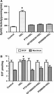
          High-density lipoprotein (HDL) has a significant cardioprotective effects. HDL induces cyclooxygenase-2 (COX-2) expression and prostacyclin I-2 (PGI-2) release in vascular endothelial cells, which contributes to its anti-atherogenic effects. However, the underlying mechanisms are not fully understood. In the present study, we observed that HDL-stimulated COX-2 expression and PGI-2 production in human umbilical vein endothelial cells (HUVECs) in a time- and dose-dependent manner. These effects triggered by HDL were inhibited by pertussis toxin (PTX), protein kinase C (PKC) inhibitor GF109203X, and ERK inhibitor PD98059, suggesting that Gαi/Gαo-coupled GPCR, PKC, and ERK pathways are involved in HDL-induced COX-2/PGI-2 activation. More importantly, we found that silencing of sphingosine kinase 2 (SphK-2) also blocked HDL-induced COX-2/PGI-2 activation. In addition, HDL-activated SphK-2 phosphorylation accompanied by increased S1P level in the nucleus. Our ChIP data demonstrated that SphK-2 is associated with CREB at the COX-2 promoter region. Collectively, these results indicate that HDL induces COX-2 expression and PGI-2 release in endothelial cells through activation of PKC, ERK1/2, and SphK-2 pathways. These findings implicate a novel mechanism underlying anti-atherothrombotic effects of HDL.

          Related collections

          Most cited references30

          • Record: found
          • Abstract: found
          • Article: not found

          Endothelial and antithrombotic actions of HDL.

          It is well recognized that high-density lipoprotein (HDL)-cholesterol is antiatherogenic and serves a role in mediating cholesterol efflux from cells. However, HDL has multiple additional endothelial and antithrombotic actions that may also afford cardiovascular protection. HDL promotes the production of the atheroprotective signaling molecule nitric oxide (NO) by upregulating endothelial NO synthase (eNOS) expression, by maintaining the lipid environment in caveolae where eNOS is colocalized with partner signaling molecules, and by stimulating eNOS as a result of kinase cascade activation by the high-affinity HDL receptor scavenger receptor class B type I (SR-BI). HDL also protects endothelial cells from apoptosis and promotes their growth and their migration via SR-BI-initiated signaling. As importantly, there is evidence of a variety of mechanisms by which HDL is antithrombotic and thereby protective against arterial and venous thrombosis, including through the activation of prostacyclin synthesis. The antithrombotic properties may also be related to the abilities of HDL to attenuate the expression of tissue factor and selectins, to downregulate thrombin generation via the protein C pathway, and to directly and indirectly blunt platelet activation. Thus, in addition to its cholesterol-transporting properties, HDL favorably regulates endothelial cell phenotype and reduces the risk of thrombosis. With further investigation and resulting greater depth of understanding, these mechanisms may be harnessed to provide new prophylactic and therapeutic strategies to combat atherosclerosis and thrombotic disorders.
            Bookmark
            • Record: found
            • Abstract: found
            • Article: not found

            Erythrocytes store and release sphingosine 1-phosphate in blood.

            The blood constituent sphingosine 1-phosphate (S1P) is a specific ligand for five G-protein-coupled receptors designated S1P(1-5). Expression of the S1P1 receptor on lymphocytes is required for their exit from secondary lymphoid organs, suggesting that S1P serves as a stimulus for maintaining lymphocyte circulation in blood. Despite its potential role in immune surveillance, the regulatory system that controls blood S1P levels is not well understood. This report reveals that erythrocytes constitute a buffer system for S1P in blood. They efficiently incorporated and stored S1P, and protected it from cellular degradation. They also released S1P into plasma, but not into other serum-free media, indicating that S1P release was controlled by a plasma factor. Erythrocytes did not generate S1P since an increase in plasma S1P levels was always accompanied by a decrease in cellular S1P levels. Thrombocytes that were reported to generate and release S1P after activation did not contribute to the observed S1P release in blood. The amount of erythrocytes as well as the proportion of plasma in the medium determined the magnitude of S1P release. Adoptively transferred S1P-loaded and unloaded mouse erythrocytes displayed a normal life span and similar S1P levels 24 h after recovery, indicating that S1P incorporation and release are dynamically regulated in vivo.
              Bookmark
              • Record: found
              • Abstract: not found
              • Article: not found

              Functions of the multifaceted family of sphingosine kinases and some close relatives.

                Bookmark

                Author and article information

                Contributors
                xslyxry@163.com
                +86-734-8281586 , +86-734-8281586 , ghyi6108@163.com
                Journal
                Mol Cell Biochem
                Mol. Cell. Biochem
                Molecular and Cellular Biochemistry
                Springer US (Boston )
                0300-8177
                1573-4919
                3 January 2014
                3 January 2014
                2014
                : 389
                : 197-207
                Affiliations
                [ ]You Country People’s Hospital, Zhuzhou, 412300 Hunan China
                [ ]Institute of Cardiovascular Disease Research, Key Laboratory for Atherosclerology of Hunan Province, University of South China, No. 28, Changsheng Road, Hengyang, 421001 Hunan China
                Article
                1941
                10.1007/s11010-013-1941-y
                3950625
                24385109
                f43fcd4f-9c19-49c2-985e-d297b1e96cfa
                © The Author(s) 2014

                Open AccessThis article is distributed under the terms of the Creative Commons Attribution License which permits any use, distribution, and reproduction in any medium, provided the original author(s) and the source are credited.

                History
                : 29 August 2013
                : 18 December 2013
                Categories
                Article
                Custom metadata
                © Springer Science+Business Media New York 2014

                Biochemistry
                high-density lipoprotein,sphingosine kinase,sphingosine 1-phosphate,cyclooxygenase-2,protein kinase c

                Comments

                Comment on this article