Inviting an author to review:
Find an author and click ‘Invite to review selected article’ near their name.
Search for authorsSearch for similar articles
17
views
0
recommends
+1 Recommend
0 collections
    0
    shares
      • Record: found
      • Abstract: found
      • Article: found
      Is Open Access

      Promising Low-Toxicity of Viologen-Phosphorus Dendrimers against Embryonic Mouse Hippocampal Cells

      research-article

      Read this article at

      Bookmark
          There is no author summary for this article yet. Authors can add summaries to their articles on ScienceOpen to make them more accessible to a non-specialist audience.

          Abstract

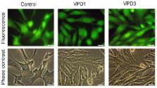
          A new class of viologen-phosphorus dendrimers (VPDs) has been recently shown to possess the ability to inhibit neurodegenerative processes in vitro. Nevertheless, in the Central Nervous Systems domain, there is little information on their impact on cell functions, especially on neuronal cells. In this work, we examined the influence of two VPD (VPD1 and VPD3) of zero generation (G0) on murine hippocampal cell line (named mHippoE-18). Extended analyses of cell responses to these nanomolecules comprised cytotoxicity test, reactive oxygen species (ROS) generation studies, mitochondrial membrane potential (ΔΨm) assay, cell death detection, cell morphology assessment, cell cycle studies, as well as measurements of catalase (CAT) activity and glutathione (GSH) level. The results indicate that VPD1 is more toxic than VPD3. However, these two tested dendrimers did not cause a strong cellular response, and induced a low level of apoptosis. Interestingly, VPD1 and VPD3 treatment led to a small decline in ROS level compared to untreated cells, which correlated with slightly increased catalase activity. This result indicates that the VPDs can indirectly lower the level of ROS in cells. Summarising, low-cytotoxicity on mHippoE-18 cells together with their ability to quench ROS, make the VPDs very promising nanodevices for future applications in the biomedical field as nanocarriers and/or drugs per se.

          Related collections

          Most cited references56

          • Record: found
          • Abstract: found
          • Article: not found

          Role of reactive oxygen species (ROS) in apoptosis induction.

          Reactive oxygen species (ROS) and mitochondria play an important role in apoptosis induction under both physiologic and pathologic conditions. Interestingly, mitochondria are both source and target of ROS. Cytochrome c release from mitochondria, that triggers caspase activation, appears to be largely mediated by direct or indirect ROS action. On the other hand, ROS have also anti-apoptotic effects. This review focuses on the role of ROS in the regulation of apoptosis, especially in inflammatory cells.
            Bookmark
            • Record: found
            • Abstract: found
            • Article: found
            Is Open Access

            A simple technique for quantifying apoptosis in 96-well plates

            Background Analyzing apoptosis has been an integral component of many biological studies. However, currently available methods for quantifying apoptosis have various limitations including multiple, sometimes cell-damaging steps, the inability to quantify live, necrotic and apoptotic cells at the same time, and non-specific detection (i.e. "false positive"). To overcome the shortcomings of current methods that quantify apoptosis in vitro and to take advantage of the 96-well plate format, we present here a modified ethidium bromide and acridine orange (EB/AO) staining assay, which may be performed entirely in a 96-well plate. Our method combines the advantages of the 96-well format and the conventional EB/AO method for apoptotic quantification. Results We compared our method and the conventional EB/AO method for quantifying apoptosis of suspension cells (Jurkat) and adherent cells (A375) under normal growth and apoptosis-inducing conditions. We found that our new EB/AO method achieved quantification results comparable to those produced using the conventional EB/AO method for both suspension and adherent cells. Conclusion By eliminating the detaching and washing steps, our method drastically reduces the time needed to perform the test, minimizes damage to adherent cells, and decreases the possibility of losing floating cells. Overall, our method is an improvement over the currently available techniques especially for adherent cells.
              Bookmark
              • Record: found
              • Abstract: found
              • Article: not found

              JC-1, but not DiOC6(3) or rhodamine 123, is a reliable fluorescent probe to assess delta psi changes in intact cells: implications for studies on mitochondrial functionality during apoptosis.

              The sensitivity and specificity of three fluorescent probes used for cytofluorimetric analysis of mitochondrial membrane potential (delta psi) were studied in the U937 human cell line. First, the role of plasmamembrane in influencing the binding of the probes to mitochondria has been investigated. The depolarization of plasmamembrane with high doses of extracellular KCl had no immediate effects on the loading of JC-1, DiOC6(3) and rhodamine 123 (R123). However, after a few hours of culture in the presence of KCl, significant changes were observed only in cells stained with DiOC6(3). Second, a comparative study was performed concerning the effects of agents capable of collapsing deltapsi. While adding FCCP to cell cultures resulted in consistent changes in the fluorescence emission of both JC-1 and DiOC6(3) - but not of R123 - only cells stained with JC-1 responded to valinomycin. On the whole, our data indicate that JC-1 is a reliable probe for analyzing delta psi changes with flow cytometry, while the others show a lower sensitivity (R123), or a non-coherent behaviour, due to a high sensitivity to changes in plasmamembrane potential [DiOC6(3)]. These data cast some doubts on those studies that, using fluorescent probes that have a low sensitivity to delta psi, hypothesized that the fall in delta psi is one of the early events, if not one of the main causes, of apoptosis.
                Bookmark

                Author and article information

                Journal
                Molecules
                Molecules
                molecules
                Molecules
                MDPI
                1420-3049
                30 September 2013
                October 2013
                : 18
                : 10
                : 12222-12240
                Affiliations
                [1 ]Department of General Biophysics, Faculty of Biology and Environmental Protection, University of Lodz, Pomorska 141/143, Lodz 90236, Poland
                [2 ]Laboratoire de Chimie de Coordination CNRS, 205 Route de Narbonne, Toulouse 31077, France
                [3 ]Laboratoire de Chimie et de Biochimie Pharmacologiques et Toxicologique, Université Paris Descartes, PRES Sorbonne Paris Cité, CNRS UMR 860, 45, rue des Saints Pères, Paris 75006, France
                [4 ]Institute of Nanomaterials and Nanotechnology (INANOTECH)-MAScIR (Moroccan Foundation for Advanced Science, Innovation and Research), ENSET, Avenue de l’Armée Royale, Madinat El Irfane, Rabat 10100, Morocco
                Author notes
                [* ]Author to whom correspondence should be addressed; E-Mail: aklajn@ 123456biol.uni.lodz.pl ; Tel.: +48-42-635-44-29; Fax: +48-42-635-44-74.
                Article
                molecules-18-12222
                10.3390/molecules181012222
                6270227
                24084024
                f49b548b-0cd1-4ae1-90a7-a64d5f71b213
                © 2013 by the authors; licensee MDPI, Basel, Switzerland.

                This article is an open access article distributed under the terms and conditions of the Creative Commons Attribution license ( http://creativecommons.org/licenses/by/3.0/).

                History
                : 12 August 2013
                : 24 September 2013
                : 24 September 2013
                Categories
                Article

                apoptosis,cytotoxicity,hippocampal cell line,ros,viologen-phosphorus dendrimers

                Comments

                Comment on this article