53
views
0
recommends
+1 Recommend
0 collections
    0
    shares
      • Record: found
      • Abstract: found
      • Article: not found

      Ataxin-3 Is a Multivalent Ligand for the Parkin Ubl Domain

      research-article

      Read this article at

      ScienceOpenPublisherPMC
      Bookmark
          There is no author summary for this article yet. Authors can add summaries to their articles on ScienceOpen to make them more accessible to a non-specialist audience.

          Abstract

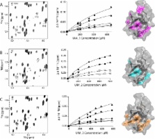
          The ubiquitin signaling pathway consists of hundreds of enzymes that are tightly regulated for the maintenance of cell homeostasis. Parkin is an E3 ubiquitin ligase responsible for conjugating ubiquitin onto a substrate protein, which itself can be ubiquitinated. Ataxin-3 performs the opposing function as a deubiquitinating enzyme that can remove ubiquitin from parkin. In this work, we have identified the mechanism of interaction between the ubiquitin-like (Ubl) domain from parkin and three C-terminal ubiquitin-interacting motifs (UIMs) in ataxin-3. 1H– 15N heteronuclear single-quantum coherence titration experiments revealed that there are weak direct interactions between all three individual UIM regions of ataxin-3 and the Ubl domain. Each UIM utilizes the exposed β-grasp surface of the Ubl domain centered around the I44 patch that did not vary in the residues involved or the surface size as a function of the number of ataxin-3 UIMs involved. Further, the apparent dissociation constant for ataxin-3 decreased as a function of the number of UIM regions used in experiments. A global multisite fit of the nuclear magnetic resonance titration data, based on three identical binding ligands, resulted in a K D of 669 ± 62 μM for each site. Our observations support a multivalent ligand binding mechanism employed by the parkin Ubl domain to recruit multiple UIM regions in ataxin-3 and provide insight into how these two proteins function together in ubiquitination–deubiquitination pathways.

          Related collections

          Most cited references32

          • Record: found
          • Abstract: found
          • Article: not found

          Structure of parkin reveals mechanisms for ubiquitin ligase activation.

          Mutations in the PARK2 (parkin) gene are responsible for an autosomal recessive form of Parkinson's disease. The parkin protein is a RING-in-between-RING E3 ubiquitin ligase that exhibits low basal activity. We describe the crystal structure of full-length rat parkin. The structure shows parkin in an autoinhibited state and provides insight into how it is activated. RING0 occludes the ubiquitin acceptor site Cys(431) in RING2, whereas a repressor element of parkin binds RING1 and blocks its E2-binding site. Mutations that disrupted these inhibitory interactions activated parkin both in vitro and in cells. Parkin is neuroprotective, and these findings may provide a structural and mechanistic framework for enhancing parkin activity.
            Bookmark
            • Record: found
            • Abstract: found
            • Article: not found

            Autoregulation of Parkin activity through its ubiquitin-like domain.

            Parkin is an E3-ubiquitin ligase belonging to the RBR (RING-InBetweenRING-RING family), and is involved in the neurodegenerative disorder Parkinson's disease. Autosomal recessive juvenile Parkinsonism, which is one of the most common familial forms of the disease, is directly linked to mutations in the parkin gene. However, the molecular mechanisms of Parkin dysfunction in the disease state remain to be established. We now demonstrate that the ubiquitin-like domain of Parkin functions to inhibit its autoubiquitination. Moreover pathogenic Parkin mutations disrupt this autoinhibition, resulting in a constitutively active molecule. In addition, we show that the mechanism of autoregulation involves ubiquitin binding by a C-terminal region of Parkin. Our observations provide important molecular insights into the underlying basis of Parkinson's disease, and in the regulation of RBR E3-ligase activity.
              Bookmark
              • Record: found
              • Abstract: found
              • Article: not found

              Parkin ubiquitinates the alpha-synuclein-interacting protein, synphilin-1: implications for Lewy-body formation in Parkinson disease.

              Parkinson disease is a common neurodegenerative disorder characterized by the loss of dopaminergic neurons and the presence of intracytoplasmic-ubiquitinated inclusions (Lewy bodies). Mutations in alpha-synuclein (A53T, A30P) and parkin cause familial Parkinson disease. Both these proteins are found in Lewy bodies. The absence of Lewy bodies in patients with parkin mutations suggests that parkin might be required for the formation of Lewy bodies. Here we show that parkin interacts with and ubiquitinates the alpha-synuclein-interacting protein, synphilin-1. Co-expression of alpha-synuclein, synphilin-1 and parkin result in the formation of Lewy-body-like ubiquitin-positive cytosolic inclusions. We further show that familial-linked mutations in parkin disrupt the ubiquitination of synphilin-1 and the formation of the ubiquitin-positive inclusions. These results provide a molecular basis for the ubiquitination of Lewy-body-associated proteins and link parkin and alpha-synuclein in a common pathogenic mechanism through their interaction with synphilin-1.
                Bookmark

                Author and article information

                Journal
                Biochemistry
                Biochemistry
                bi
                bichaw
                Biochemistry
                American Chemical Society
                0006-2960
                1520-4995
                24 September 2013
                22 October 2013
                : 52
                : 42
                : 7369-7376
                Affiliations
                Department of Biochemistry, Schulich School of Medicine and Dentistry, University of Western Ontario , London, Ontario, Canada N6A 5C1
                Author notes
                [* ]Department of Biochemistry, University of Western Ontario, London, Ontario N6A 5C1, Canada. E-mail: gshaw1@ 123456uwo.ca . Phone: (519) 661-4021. Fax: (519) 661-3175.
                Article
                10.1021/bi400780v
                3807529
                24063750
                f782a68f-ae69-4bf8-81e1-2382bd0b9a60
                Copyright © 2013 American Chemical Society
                History
                : 17 June 2013
                : 23 September 2013
                Categories
                Article
                Custom metadata
                bi400780v
                bi-2013-00780v

                Biochemistry
                Biochemistry

                Comments

                Comment on this article