40
views
0
recommends
+1 Recommend
0 collections
    0
    shares
      • Record: found
      • Abstract: found
      • Article: found
      Is Open Access

      Asiatic acid attenuates methamphetamine-induced neuroinflammation and neurotoxicity through blocking of NF-kB/STAT3/ERK and mitochondria-mediated apoptosis pathway

      research-article

      Read this article at

      Bookmark
          There is no author summary for this article yet. Authors can add summaries to their articles on ScienceOpen to make them more accessible to a non-specialist audience.

          Abstract

          Background

          Methamphetamine (METH) is a commonly abused drug that may result in neurotoxic effects. Recent studies have suggested that involvement of neuroinflammatory processes in brain dysfunction is induced by misuse of this drug. However, the mechanism underlying METH-induced inflammation and neurotoxicity in neurons is still unclear. In this study, we investigated whether asiatic acid (AA) effected METH-mediated neuroinflammation and neurotoxicity in dopaminergic neuronal cells. And we further determined whether the effect involved in the nuclear factor kappa-light-chain-enhancer of activated B cells (NF-κB) and signal transducer and activator of transcription (STAT)3 and extracellular signal-regulated kinase (ERK) pathway.

          Methods

          We used the human dopaminergic neuroblastoma SH-SY5Y cell line, murine microglial BV2 cell line, and primary culture of rat embryo mesencephalic neurons. Pro-inflammatory cytokine production was monitored by ELISA and RT/real-time PCR. The cell cycle distribution and mitochondrial membrane integrity was analyzed by flow cytometry. We used immunoblotting, DNA-binding activity, and immunofluorescence staining to analyze the effect of AA on activation of the NF-κB, STAT3, MAPK-ERK, and apoptosis signaling pathways.

          Results

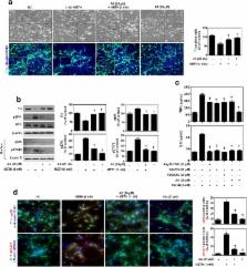
          METH induced TNF receptor (TNFR) expression and led to morphological changes of cells. Additionally, this drug increased pro-inflammatory cytokine (TNFα and IL-6) expression. AA significantly suppressed METH-induced TNFR expression in concentration dependent. Increased secretion of TNFα and IL-6 was inhibited in METH-stimulated neuronal cells by AA administration. AA showed significant protection against METH-induced translocation of NF-κB/STAT3 and ERK phosphorylation. AA inhibited METH-induced proteolytic fragmentation of caspase-3 and PARP. The pro-apoptotic protein Bax was significantly decreased, while the anti-apoptotic protein Bcl-xL was increased by AA treatment in METH-stimulated cells. A similar protective effect of AA on mitochondrial membrane integrity was also confirmed by flow cytometry and immunofluorescence staining.

          Conclusions

          Based on the literatures and our findings, AA is a promising candidate for an anti-neurotoxic agent, and it can potentially be used for the prevention and treatment of various neurological disorders.

          Electronic supplementary material

          The online version of this article (10.1186/s12974-017-1009-0) contains supplementary material, which is available to authorized users.

          Related collections

          Most cited references42

          • Record: found
          • Abstract: found
          • Article: found
          Is Open Access

          Neuropeptides and Microglial Activation in Inflammation, Pain, and Neurodegenerative Diseases

          Microglial cells are responsible for immune surveillance within the CNS. They respond to noxious stimuli by releasing inflammatory mediators and mounting an effective inflammatory response. This is followed by release of anti-inflammatory mediators and resolution of the inflammatory response. Alterations to this delicate process may lead to tissue damage, neuroinflammation, and neurodegeneration. Chronic pain, such as inflammatory or neuropathic pain, is accompanied by neuroimmune activation, and the role of glial cells in the initiation and maintenance of chronic pain has been the subject of increasing research over the last two decades. Neuropeptides are small amino acidic molecules with the ability to regulate neuronal activity and thereby affect various functions such as thermoregulation, reproductive behavior, food and water intake, and circadian rhythms. Neuropeptides can also affect inflammatory responses and pain sensitivity by modulating the activity of glial cells. The last decade has witnessed growing interest in the study of microglial activation and its modulation by neuropeptides in the hope of developing new therapeutics for treating neurodegenerative diseases and chronic pain. This review summarizes the current literature on the way in which several neuropeptides modulate microglial activity and response to tissue damage and how this modulation may affect pain sensitivity.
            Bookmark
            • Record: found
            • Abstract: found
            • Article: not found

            Annexin A1: a central player in the anti-inflammatory and neuroprotective role of microglia.

            The brain microenvironment is continuously monitored by microglia with the detection of apoptotic cells or pathogens being rapidly followed by their phagocytosis to prevent inflammatory responses. The protein annexin A1 (ANXA1) is key to the phagocytosis of apoptotic leukocytes during peripheral inflammatory resolution, but the pathophysiological significance of its expression in the CNS that is restricted almost exclusively to microglia is unclear. In this study, we test the hypothesis that ANXA1 is important in the microglial clearance of apoptotic neurons in both noninflammatory and inflammatory conditions. We have identified ANXA1 to be sparingly expressed in microglia of normally aged human brains and to be more strongly expressed in Alzheimer's disease. Using an in vitro model comprising microglial and neuronal cell lines, as well as primary microglia from wild-type and ANXA1 null mice, we have identified two distinct roles for microglial ANXA1: 1) controlling the noninflammatory phagocytosis of apoptotic neurons and 2) promoting resolution of inflammatory microglial activation. In particular, we showed that microglial-derived ANXA1 targets apoptotic neurons, serving as both an "eat me" signal and a bridge between phosphatidylserine on the dying cell and formyl peptide receptor 2 on the phagocytosing microglia. Moreover, inflammatory activation of microglia impairs their ability to discriminate between apoptotic and nonapoptotic cells, an ability restored by exogenous ANXA1. We thus show that ANXA1 is fundamental for brain homeostasis, and we suggest that ANXA1 and its peptidomimetics can be novel therapeutic targets in neuroinflammation.
              Bookmark
              • Record: found
              • Abstract: found
              • Article: found
              Is Open Access

              Phytochemicals That Regulate Neurodegenerative Disease by Targeting Neurotrophins: A Comprehensive Review

              Alzheimer's disease (AD), characterized by progressive dementia and deterioration of cognitive function, is an unsolved social and medical problem. Age, nutrition, and toxins are the most common causes of AD. However, currently no credible treatment is available for AD. Traditional herbs and phytochemicals may delay its onset and slow its progression and also allow recovery by targeting multiple pathological causes by antioxidative, anti-inflammatory, and antiamyloidogenic properties. They also regulate mitochondrial stress, apoptotic factors, free radical scavenging system, and neurotrophic factors. Neurotrophins such as BDNF, NGF, NT3, and NT4/5 play a vital role in neuronal and nonneuronal responses to AD. Neurotrophins depletion accelerates the progression of AD and therefore, replacing such neurotrophins may be a potential treatment for neurodegenerative disease. Here, we review the phytochemicals that mediate the signaling pathways involved in neuroprotection specifically neurotrophin-mediated activation of Trk receptors and members of p75NTR superfamily. We focus on representative phenolic derivatives, iridoid glycosides, terpenoids, alkaloids, and steroidal saponins as regulators of neurotrophin-mediated neuroprotection. Although these phytochemicals have attracted attention owing to their in vitro neurotrophin potentiating activity, their in vivo and clinical efficacy trials has yet to be established. Therefore, further research is necessary to prove the neuroprotective effects in preclinical models and in humans.
                Bookmark

                Author and article information

                Contributors
                (+82)53-580-6653 , bdpark@kmu.ac.kr
                Journal
                J Neuroinflammation
                J Neuroinflammation
                Journal of Neuroinflammation
                BioMed Central (London )
                1742-2094
                11 December 2017
                11 December 2017
                2017
                : 14
                : 240
                Affiliations
                [1 ]ISNI 0000 0001 0669 3109, GRID grid.412091.f, College of Pharmacy, , Keimyung University, ; 1095 Dalgubeoldaero, Dalseo-Gu, Daegu, 42601 Republic of Korea
                [2 ]ISNI 0000 0001 0669 3109, GRID grid.412091.f, Department of Pharmacology, School of Medicine, , Keimyung University, ; 1095 Dalgubeoldaero, Dalseo-Gu, Daegu, 42601 Republic of Korea
                Article
                1009
                10.1186/s12974-017-1009-0
                5725763
                29228978
                f7dbe435-cf3c-4c03-ab76-45798b8e4c2d
                © The Author(s). 2017

                Open AccessThis article is distributed under the terms of the Creative Commons Attribution 4.0 International License ( http://creativecommons.org/licenses/by/4.0/), which permits unrestricted use, distribution, and reproduction in any medium, provided you give appropriate credit to the original author(s) and the source, provide a link to the Creative Commons license, and indicate if changes were made. The Creative Commons Public Domain Dedication waiver ( http://creativecommons.org/publicdomain/zero/1.0/) applies to the data made available in this article, unless otherwise stated.

                History
                : 20 April 2017
                : 22 November 2017
                Categories
                Research
                Custom metadata
                © The Author(s) 2017

                Neurosciences
                methamphetamine,asiatic acid,neuroinflammation,neurotoxicity,nf-κb,stat3,erk
                Neurosciences
                methamphetamine, asiatic acid, neuroinflammation, neurotoxicity, nf-κb, stat3, erk

                Comments

                Comment on this article