- Record: found
- Abstract: found
- Article: found
Atlantoaxial Subluxation Associated with Os Odontoideum Fused to the Anterior Arch of the Atlas: A Case Report
brief-report
Read this article at
There is no author summary for this article yet. Authors can add summaries to their articles on ScienceOpen to make them more accessible to a non-specialist audience.
Abstract
There are various anomalies in the craniocervical junction due to the complex nature
of the cranial and cervical bone development
1
). Fusion of os odontoideum and the atlas (C1) has been very rarely reported
2
). Since the first case described by Wackenheim in 1971
3
), very few cases have been reported in the literature
4-6
). Herein, we report a case of atlantoaxial subluxation associated with the fusion
of os odontoideum and the anterior arch of the atlas. A 63-year-old male patient presented
with a 2-month history of increasing posterior neck pain, clumsiness, and difficulty
in walking. He had hyperactivity of the deep tendon reflex on both sides of the upper
extremities but no other neurological abnormalities, such as muscle atrophy and lower
cranial nerve dysfunction. He had no history of neck or head trauma. Plain radiographs
of the cervical spine revealed a separation of the dens from the axis as well as atlantoaxial
subluxation (Fig. 1A), which was reduced in the extended neck position (Fig. 1B).
T2-weighted magnetic resonance imaging (MRI) revealed areas of hyperintensity in the
spinal cord at the level between the body of the axis and the posterior arch of the
atlas (Fig. 1C). Computed tomography (CT) myelography revealed fusion of the apical
segment of the dens and the anterior arch of C1 and narrowing of the spinal canal
between the basal dens and the posterior arch (Fig. 2A and D-E). Other anomalous conditions
were not identified in the atlantooccipital and atlantoaxial joints (Fig. 2B-C), and
there was no additional anomaly in the subaxial cervical spine. In this case with
progressive myelopathy, atlantoaxial arthrodesis via a posterior approach was planned
as it was considered to be reasonable. The operation was performed using the Magerl
and Brooks technique (Fig. 3), after which the patient's motor impairment improved
over time, with slight numbness in the upper extremities.
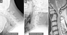
Figure 1.
Preoperative plain radiography and magnetic resonance imaging (MRI).
Plain radiographs showing that atlantoaxial subluxation is reduced in the extension
position (A–B). MRI showing high-signal intensity within the spinal cord at the level
between the basal dental segment and the posterior arch of C1 (C).
Figure 2.
Preoperative computed tomography (CT) myelography.
CT showing the anterior arch of C1 fused with the apical segment of the dens separated
from the basal segment of the dens (A). Sagittal reconstruction CT showing the right
and left C0–C1–C2 articulations (B–C). Atlantoaxial dislocation in the flexion position
(D) is reduced in the neutral position (E).
Figure 3.
Atlantoaxial fusion surgery.
Atlantoaxial arthrodesis was performed using the Magerl and Brooks technique.
In general, the odontoid process separates from the anterior part of the atlas and
caudally migrates to fuse with the body of the axis between the 6th and 7th weeks
of gestation. After resegmentation of cervical sclerotomes, the odontoid process is
composed of the apical dental segment from the caudal proatlas, the basal dental segment
from the first cervical sclerotomes, and the body of the axis from the second cervical
sclerotomes
7
) (Fig. 4). As a result of the complex processes involved in the embryological development
of the cervical spine (especially segmentation and resegmentation), various anomalies
can occur in the occipitocervical region
1
).
Figure 4.
Embryology of the axis.
The odontoid process comprises the apical dental segment from the caudal proatlas,
the basal dental segment from the first cervical sclerotome, and the body of the axis
from the second cervical sclerotome.
PA: proatlas, CSo: cervical somite, CSc: cervical sclerotome
Previous reports have demonstrated that os odontoideum, an anomaly of the axis, has
an occurrence rate of 0.7%-0.8%
8
,
9
), and 32%-44% patients with os odontoideum have progressive myelopathy
10
,
11
). However, only 10 cases of os odontoideum fused with the anterior arch of the atlas
have been reported thus far, among which only 2 (20%) had progressive myelopathy with
atlantoaxial dislocation
2
,
12
). There are limited data on the differences in the mechanical properties of the atlantoaxial
joint between the two anomalous conditions. However, given the occurrence rate of
neurological symptoms, there would be no significant differences in the mechanical
instability of the atlantoaxial joint between patients with os odontoideum and those
with fused anterior arch of the atlas. In our case, the patient developed cervical
myelopathy in his 60s without a history of major trauma. We speculated that the cumulative
micromechanical stress of daily living with the incomplete bony structures might have
caused the atlantoaxial instability.
In this study, we have described a rare case of a 63-year-old male patient with atlantoaxial
subluxation and progressive myelopathy associated with the fusion of os odontoideum
and the anterior arch of the atlas.
Conflicts of Interest: The authors declare that there are no relevant conflicts of
interest.
Sources of Funding: None.
Author Contributions: Drs. Fujiwara and Akeda contributed equally to this manuscript.
All of the authors had read, reviewed, and approved the manuscript.
Ethical Approval: This article does not contain any studies with human participants
performed by any of the authors.
Informed Consent: Written informed consent was obtained from the patient for publication
of this case report and any accompanying images.
Related collections
Author and article information
Comments
Comment on this article
scite_