17
views
0
recommends
+1 Recommend
0 collections
    0
    shares
      • Record: found
      • Abstract: found
      • Article: found
      Is Open Access

      Proteomic and Ultrastructural Analysis of Cellulite—New Findings on an Old Topic

      research-article

      Read this article at

      Bookmark
          There is no author summary for this article yet. Authors can add summaries to their articles on ScienceOpen to make them more accessible to a non-specialist audience.

          Abstract

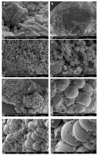
          Background: Cellulite is a condition in which the skin has a dimpled lumpy appearance. The main causes of cellulite development, studied until now, comprehends modified sensitivity to estrogens, the damage of microvasculature present among dermis and hypodermis. The differences of adipose tissue architecture between male and female might make female more susceptible to cellulite. Adipose tissue is seen to be deeply modified during cellulite development. Our study tried to understand the overall features within and surrounding cellulite to apply the best therapeutic approach. Methods: Samples of gluteal femoral area were collected from cadavers and women who had undergone surgical treatment to remove orange peel characteristics on the skin. Samples from cadavers were employed for an accurate study of cellulite using magnetic resonance imaging at 7 Tesla and for light microscopy. Specimens from patients were employed for the proteomic analysis, which was performed using high resolution mass spectroscopy (MS). Stromal vascular fraction (SVF) was obtained from the samples, which was studied using MS and flow cytometry. Results: light and electron microscopy of the cellulite affected area showed a morphology completely different from the other usual adipose depots. In cellulite affected tissues, sweat glands associated with adipocytes were found. In particular, there were vesicles in the extracellular matrix, indicating a crosstalk between the two different components. Proteomic analysis showed that adipose tissue affected by cellulite is characterized by high degree of oxidative stress and by remodeling phenomena. Conclusions: The novel aspects of this study are the peculiar morphology of adipose tissue affected by cellulite, which could influence the surgical procedures finalized to the reduction of dimpling, based on the collagen fibers cutting. The second novel aspect is the role played by the mesenchymal stem cells isolated from stromal vascular fraction of adipose tissue affected by cellulite.

          Related collections

          Most cited references21

          • Record: found
          • Abstract: found
          • Article: not found

          Treatment of cellulite: Part I. Pathophysiology.

          Cellulite is a topographic skin change that is nearly ubiquitous in postpubertal women. Treatment remains elusive. The various treatments currently available are only partially or temporarily effective. Newer therapeutic modalities continue to evolve without much understanding of the complex nature of cellulite. The successful treatment of cellulite will ultimately depend upon our understanding of the pathophysiology of cellulite adipose tissue. Part I of this two-part series on cellulite reviews how the concept and perception of cellulite has evolved over time and its proposed etiologies. The article also focuses on the physiology of human adipose tissue, particularly regarding cellulite. Copyright 2009 American Academy of Dermatology, Inc. Published by Mosby, Inc. All rights reserved.
            Bookmark
            • Record: found
            • Abstract: found
            • Article: not found

            Cellulite: nature and aetiopathogenesis.

            Only a limited number of studies on cellulite have been published in the international literature and many of them reach somewhat antithetical conclusions. Consequently, it is not yet possible to reconcile the extreme differences of opinion which have lingered on for years concerning the nature of this disorder, as well as its origin and even the most basic aspects of its histopathological classification. It does not even have a recognized name: in fact, the term 'cellulitis' is used in scientific English to indicate a spreading gangrenous infection of the subcutaneous cellular tissue. The other terms used from time to time [panniculitis, lipodystrophy, edematofibrosclerotic panniculitis (EFP), liposclerosis, lipoedema, etc.] have quite different morphological and pathogenetic connotations in general. Over the last few decades, three major conflicting theories have emerged in relation to the ethiopathogenesis of cellulite. These indicate, respectively, the following causes: 1. Oedema caused by excessive hydrophilia of the intercellular matrix. 2. A homeostatic alteration on a regional microcirculatory level; this pathogenetic theory is summarized in a synthetic and self-explanatory denomination: EFP. 3. A peculiar anatomical conformation of the subcutaneous tissue of women, different from male morphology. These theories must all now be updated in the light of recent advances on the sophisticated and composite physiopathology of the adipose organ - which acts not only as a control device which regulates the systematic equilibrium of energy and modulates the food intake and the metabolism of other tissue substrate through a multiple glandular secretion of hormones and parahormones.
              Bookmark
              • Record: found
              • Abstract: found
              • Article: not found

              Anti-fibrosclerotic effects of shock wave therapy in lipedema and cellulite.

              In vivo measurements in 26 female patients with lipedema and cellulite parameters were carried out before and after therapy by means of complex physical decongestive therapy (CPDT) including manual lymph drainage and compression as main components and/or shock wave therapy (SWT). Oxidative stress parameters of blood serum and biomechanic skin properties/smoothening of dermis and hypodermis surface were evaluated. Oxidative stress in lipedema and cellulite was demonstrated by increased serum concentrations of malondialdehyde (MDA) and plasma protein carbonyls compared with healthy control persons. Both MDA and protein carbonyls in blood plasma decreased after serial shock wave application and CPDT. The SWT itself and CPDT itself lead to MDA release from edematous tissue into the plasma. Obviously both therapy types, SWT and CPDT, mitigate oxidative stress in lipedema and cellulite. In parallel SWT improved significantly the biomechanic skin properties leading to smoothening of dermis and hypodermis surface. Significant correlation between MDA depletion of edematous and lipid enriched dermis and improvement of mechanic skin properties was demonstrated. From these findings it is concluded, that a release of lipid peroxidation (LPO) products from edematous dermis is an important sclerosis-preventing effect of SWT and/or CPDT in lipedema and cellulite. Expression of factors stimulating angiogenesis and lymphangiogenesis such as VEGF was not induced by SWT and/or CPDT and, therefore, not involved in beneficial effects by SWT and/or CPDT.
                Bookmark

                Author and article information

                Journal
                Int J Mol Sci
                Int J Mol Sci
                ijms
                International Journal of Molecular Sciences
                MDPI
                1422-0067
                18 March 2020
                March 2020
                : 21
                : 6
                : 2077
                Affiliations
                [1 ]Department of Neurosciences, Biomedicine and Movement Sciences, Anatomy and Histology division, University of Verona, 37134 Verona, Italy
                [2 ]Accademia del Lipofilling, Research and Training Center in Regenerative Surgery, 61025 Montelabbate, Italy; zingarettin@ 123456gmail.com (N.Z.);
                [3 ]Clinic of Plastic and Reconstructive Surgery, Academic Hospital of Udine, Department of Medical Area (DAME), University of Udine, 33100 Udine, Italy
                [4 ]Master of Aesthetic Regenerative and Anti-Aging Medicine, University of Verona, 37134 Verona, Italy
                [5 ]Department of Biochemistry, University of Verona, 37134 Verona, Italy
                [6 ]Department of Sciences and Technological Innovation, University of Eastern Piedmont, 15121 Alessandria, Italy
                [7 ]Department of Informatics, University of Verona, 37134 Verona, Italy
                [8 ]Department of Reconstructive and Hand Surgery, AOU Ospedali Riuniti, 60020 Ancona, Italy
                [9 ]Veternirary Medical School, University of Camerino, 62024 Camerino, Italy
                Author notes
                Author information
                https://orcid.org/0000-0002-1575-0029
                https://orcid.org/0000-0003-2977-7828
                Article
                ijms-21-02077
                10.3390/ijms21062077
                7139738
                32197394
                f9a16b45-b7ee-4803-a0e3-2cee1c315d4f
                © 2020 by the authors.

                Licensee MDPI, Basel, Switzerland. This article is an open access article distributed under the terms and conditions of the Creative Commons Attribution (CC BY) license ( http://creativecommons.org/licenses/by/4.0/).

                History
                : 21 January 2020
                : 17 March 2020
                Categories
                Article

                Molecular biology
                adult stem cells,mesenchymal stem cell,cellulite,proteomic analysis,dermal adipose tissue

                Comments

                Comment on this article