35
views
0
recommends
+1 Recommend
0 collections
    0
    shares
      • Record: found
      • Abstract: found
      • Article: found
      Is Open Access

      Characterizing Focused-Ultrasound Mediated Drug Delivery to the Heterogeneous Primate Brain In Vivo with Acoustic Monitoring

      research-article

      Read this article at

      Bookmark
          There is no author summary for this article yet. Authors can add summaries to their articles on ScienceOpen to make them more accessible to a non-specialist audience.

          Abstract

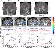
          Focused ultrasound with microbubbles has been used to noninvasively and selectively deliver pharmacological agents across the blood-brain barrier (BBB) for treating brain diseases. Acoustic cavitation monitoring could serve as an on-line tool to assess and control the treatment. While it demonstrated a strong correlation in small animals, its translation to primates remains in question due to the anatomically different and highly heterogeneous brain structures with gray and white matteras well as dense vasculature. In addition, the drug delivery efficiency and the BBB opening volume have never been shown to be predictable through cavitation monitoring in primates. This study aimed at determining how cavitation activity is correlated with the amount and concentration of gadolinium delivered through the BBB and its associated delivery efficiency as well as the BBB opening volume in non-human primates. Another important finding entails the effect of heterogeneous brain anatomy and vasculature of a primate brain, i.e., presence of large cerebral vessels, gray and white matter that will also affect the cavitation activity associated with variation of BBB opening in different tissue types, which is not typically observed in small animals. Both these new findings are critical in the primate brain and provide essential information for clinical applications.

          Related collections

          Most cited references32

          • Record: found
          • Abstract: found
          • Article: not found

          Effect of focused ultrasound applied with an ultrasound contrast agent on the tight junctional integrity of the brain microvascular endothelium.

          Previous studies have investigated a potential method for targeted drug delivery in the central nervous system that uses focused ultrasound bursts combined with an ultrasound contrast agent to temporarily disrupt the blood-brain barrier (BBB). The purpose of this work was to investigate the integrity of the tight junctions (TJs) in rat brain microvessels after this BBB disruption. Ultrasound bursts (1.5-MHz) in combination with a gas contrast agent (Optison) was applied at two locations in the brain in 25 rats to induce BBB disruption. Using immunoelectron microscopy, the distributions of the TJ-specific transmembrane proteins occludin, claudin-1, claudin-5, and of submembranous ZO-1 were examined at 1, 2, 4, 6 and 24 h after sonication. A quantitative evaluation of the protein expression was made by counting the number of immunosignals per micrometer in the junctional clefts. BBB disruption at the sonicated locations was confirmed by the leakage of i.v. administered horseradish peroxidase (HRP, m.w. 40,000 Da) and lanthanum chloride (La(3+), m.w. approximately 139 Da). Leakage of these agents was observed at 1 and 2 h and, in a few vessels, at 4 h after ultrasound application. These changes were paralleled by the apparent disintegration of the TJ complexes, as evidenced by the redistribution and loss of the immunosignals for occludin, claudin-5 and ZO-1. Claudin-1 seemed less involved. At 6 and 24 h after sonication, no HRP or lanthanum leakage was observed and the barrier function of the TJs, as indicated by the localization and density of immunosignals, appeared to be completely restored. This study provides the first direct evidence that ultrasound bursts combined with a gas contrast agent cause disassembling of the TJ molecular structure, leading to loss of the junctional barrier functions in brain microvessels. The BBB disruption appears to last up to 4 h after sonication and permits the paracellular passage of agents with molecular weights up to at least 40 kDa. These promising features can be exploited in the future development of this method that could enable the delivery of drugs, antibodies or genes to targeted locations in the brain.
            Bookmark
            • Record: found
            • Abstract: found
            • Article: not found

            Blood-brain barrier disruption with focused ultrasound enhances delivery of chemotherapeutic drugs for glioblastoma treatment.

            To demonstrate the feasibility of using focused ultrasound to enhance delivery of 1,3-bis(2-chloroethyl)-1-nitrosourea (BCNU) to glioblastomas in rats with induced tumors and determine if such an approach increases treatment efficacy. All animal experiments were approved by the animal committee and adhered to the experimental animal care guidelines. A 400-kHz focused ultrasound generator was used to transcranially disrupt the blood-brain barrier (BBB) in rat brains by delivering burst-tone ultrasound energy in the presence of microbubbles. The process was monitored in vivo by using magnetic resonance (MR) imaging. Cultured C6 glioma cells implanted in Sprague-Dawley rats were used as the tumor model. BCNU (13.5 mg/kg) was administered intravenously and its concentration in brains was quantified by using high-performance liquid chromatography. MR imaging was used to evaluate the effect of treatments longitudinally, including analysis of tumor progression and animal survival, and brain tissues were histologically examined. Methods including the two-tailed unpaired t test and the Mantel-Cox test were used for statistical analyses, with a significance level of .05. Focused ultrasound significantly enhanced the penetration of BCNU through the BBB in normal (by 340%) and tumor-implanted (by 202%) brains without causing hemorrhaging. Treatment of tumor-implanted rats with focused ultrasound alone had no beneficial effect on tumor progression or on animal survival up to 60 days. Administration of BCNU only transiently controlled tumor progression; nevertheless, relative to untreated controls, animal survival was improved by treatment with BCNU alone (increase in median survival time [IST(median)], 15.7%, P = .023). Treatment with focused ultrasound before BCNU administration controlled tumor progression (day 31: 0.05 cm(3) + or - 0.1 [standard deviation] vs 0.28 cm(3) + or - 0.1) and improved animal survival relative to untreated controls (IST(median), 85.9%, P = .0015). This study demonstrates a means of increasing localized chemotherapeutic drug delivery for brain tumor treatment and strongly supports the feasibility of this treatment in a clinical setting.
              Bookmark
              • Record: found
              • Abstract: found
              • Article: not found

              Targeted disruption of the blood-brain barrier with focused ultrasound: association with cavitation activity.

              Acoustic emission was monitored during focused ultrasound exposures in conjunction with an ultrasound contrast agent (Optison) in order to determine if cavitation activity is associated with the induction of blood-brain barrier disruption (BBBD). Thirty-four locations were sonicated (frequency: 260 kHz) at targets 10 mm deep in rabbit brain (N = 9). The sonications were applied at peak pressure amplitudes ranging from 0.11 to 0.57 MPa (burst length: 10 ms; repetition frequency of 1 Hz; duration: 20 s). Acoustic emission was recorded with a focused passive cavitation detector. This emission was recorded at each location during sonications with and without Optison. Detectable wideband acoustic emission was observed only at 0.40 and 0.57 MPa. BBBD was observed in contrast MRI after sonication at 0.29-0.57 MPa. The appearance of small regions of extravasated erythrocytes appeared to be associated with this wideband emission signal. The results thus suggest that BBBD resulting from focused ultrasound pulses in the presence of Optison can occur without indicators for inertial cavitation in vivo, wideband emission and extravasation. If inertial cavitation is not responsible for the BBBD, other ultrasound/microbubble interactions are likely the source. A significant increase in the emission signal due to Optison at the second and third harmonics of the ultrasound driving frequency was found to correlate with BBBD and might be useful as an online method to indicate when the disruption occurs.
                Bookmark

                Author and article information

                Journal
                Sci Rep
                Sci Rep
                Scientific Reports
                Nature Publishing Group
                2045-2322
                17 November 2016
                2016
                : 6
                : 37094
                Affiliations
                [1 ]Department of Biomedical Engineering, Columbia University, New York , New York, USA
                [2 ]Department of Neuroscience, Columbia University, New York , New York, USA
                [3 ]Department of Radiology, Columbia University, New York , New York, USA
                Author notes
                Article
                srep37094
                10.1038/srep37094
                5112571
                27853267
                f9e3f4c7-b0b6-4aa6-aec0-45daaca0ed12
                Copyright © 2016, The Author(s)

                This work is licensed under a Creative Commons Attribution 4.0 International License. The images or other third party material in this article are included in the article’s Creative Commons license, unless indicated otherwise in the credit line; if the material is not included under the Creative Commons license, users will need to obtain permission from the license holder to reproduce the material. To view a copy of this license, visit http://creativecommons.org/licenses/by/4.0/

                History
                : 29 June 2016
                : 25 October 2016
                Categories
                Article

                Uncategorized
                Uncategorized

                Comments

                Comment on this article