12
views
0
recommends
+1 Recommend
0 collections
    0
    shares
      • Record: found
      • Abstract: found
      • Article: found
      Is Open Access

      Isolation of intimal endothelial cells from the human thoracic aorta: Study protocol

      other

      Read this article at

      Bookmark
          There is no author summary for this article yet. Authors can add summaries to their articles on ScienceOpen to make them more accessible to a non-specialist audience.

          Abstract

          Background: Vessel endothelial cells are extensively applied to study the mechanism of atherosclerosis. Some cellular sources including human umbilical artery endothelial cells (HUAECs) and human umbilical vein endothelial cells (HUVECs) are mostly applied in the experimental studies. We described a method for isolating the human endothelial cells from the human thoracic aorta.

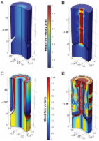
          Methods: Normal aortic samples were prepared from subjects with brain death in Masih Daneshvari Hospital. The endothelial cells were isolated using collagenase and were evaluated by the measurement of CD31 marker. Furthermore, the digestion efficacy was studied by vessel histological analysis, and the adhesion mechanism was investigated by leukocyte endothelial adhesion assay kit.

          Results and Conclusion: The isolation protocol is found as a fast and simple technique with a proper cellular load to separate the endothelial cells from the human aorta.

          Related collections

          Most cited references13

          • Record: found
          • Abstract: found
          • Article: not found

          Superoxide dismutases: role in redox signaling, vascular function, and diseases.

          Excessive reactive oxygen species Revised abstract, especially superoxide anion (O₂•-), play important roles in the pathogenesis of many cardiovascular diseases, including hypertension and atherosclerosis. Superoxide dismutases (SODs) are the major antioxidant defense systems against (O₂•-), which consist of three isoforms of SOD in mammals: the cytoplasmic Cu/ZnSOD (SOD1), the mitochondrial MnSOD (SOD2), and the extracellular Cu/ZnSOD (SOD3), all of which require catalytic metal (Cu or Mn) for their activation. Recent evidence suggests that in each subcellular location, SODs catalyze the conversion of (O₂•-), H2O2, which may participate in cell signaling. In addition, SODs play a critical role in inhibiting oxidative inactivation of nitric oxide, thereby preventing peroxynitrite formation and endothelial and mitochondrial dysfunction. The importance of each SOD isoform is further illustrated by studies from the use of genetically altered mice and viral-mediated gene transfer. Given the essential role of SODs in cardiovascular disease, the concept of antioxidant therapies, that is, reinforcement of endogenous antioxidant defenses to more effectively protect against oxidative stress, is of substantial interest. However, the clinical evidence remains controversial. In this review, we will update the role of each SOD in vascular biologies, physiologies, and pathophysiologies such as atherosclerosis, hypertension, and angiogenesis. Because of the importance of metal cofactors in the activity of SODs, we will also discuss how each SOD obtains catalytic metal in the active sites. Finally, we will discuss the development of future SOD-dependent therapeutic strategies.
            Bookmark
            • Record: found
            • Abstract: found
            • Article: not found

            Emerging Roles of Vascular Endothelium in Metabolic Homeostasis

            Our understanding of the role of the vascular endothelium has evolved over the past two decades, with the recognition that it is a dynamically regulated organ and that it plays a nodal role in a variety of physiological and pathological processes. Endothelial cells (ECs) are not only a barrier between the circulation and peripheral tissues, but also actively regulate vascular tone, blood flow and platelet function. Dysregulation of ECs contributes to pathological conditions such as vascular inflammation, atherosclerosis, hypertension, cardiomyopathy, retinopathy, neuropathy and cancer. The close anatomical relationship between vascular endothelium and highly vascularized metabolic organs/tissues suggests that the crosstalk between ECs and these organs is vital for both vascular and metabolic homeostasis. Numerous reports support that hyperlipidemia, hyperglycemia and other metabolic stresses result in endothelial dysfunction and vascular complications. However, how ECs may regulate metabolic homeostasis remains poorly understood. Emerging data suggest that the vascular endothelium plays an unexpected role in the regulation of metabolic homeostasis and that endothelial dysregulation directly contributes to the development of metabolic disorders. Here, we review recent studies about the pivotal role of ECs in glucose and lipid homeostasis. In particular, we introduce the concept that the endothelium adjusts its barrier function to control the trans-endothelial transport of fatty acids, lipoproteins, lipoprotein lipases, glucose and insulin. In addition, we summarize reports that ECs communicate with metabolic cells through EC-secreted factors and we discuss how endothelial dysregulation contributes directly to the development of obesity, insulin resistance, dyslipidemia, diabetes, cognitive defects and fatty liver disease.
              Bookmark
              • Record: found
              • Abstract: found
              • Article: found
              Is Open Access

              Biomechanical factors in the biology of aortic wall and aortic valve diseases

              The biomechanical factors that result from the haemodynamic load on the cardiovascular system are a common denominator of several vascular pathologies. Thickening and calcification of the aortic valve will lead to reduced opening and the development of left ventricular outflow obstruction, referred to as aortic valve stenosis. The most common pathology of the aorta is the formation of an aneurysm, morphologically defined as a progressive dilatation of a vessel segment by more than 50% of its normal diameter. The aortic valve is exposed to both haemodynamic forces and structural leaflet deformation as it opens and closes with each heartbeat to assure unidirectional flow from the left ventricle to the aorta. The arterial pressure is translated into tension-dominated mechanical wall stress in the aorta. In addition, stress and strain are related through the aortic stiffness. Furthermore, blood flow over the valvular and vascular endothelial layer induces wall shear stress. Several pathophysiological processes of aortic valve stenosis and aortic aneurysms, such as macromolecule transport, gene expression alterations, cell death pathways, calcification, inflammation, and neoangiogenesis directly depend on biomechanical factors.
                Bookmark

                Author and article information

                Journal
                Med J Islam Repub Iran
                Med J Islam Repub Iran
                MJIRI
                Med J Islam Repub Iran
                Medical Journal of the Islamic Republic of Iran
                Iran University of Medical Sciences
                1016-1430
                2251-6840
                2019
                04 June 2019
                : 33
                : 51
                Affiliations
                1 Biochemistry Department, Iran University of Medical Sciences, Tehran, Iran
                2 Shahid Beheshti University of Medical Sciences, Masih Daneshvari Hospital, Tehran, Iran
                3 Department of Cellular and Molecular biology, Faculty of Advanced Science and Technology, Tehran Medical Sciences, Islamic Azad University, Tehran, Iran
                4 Cellular and Molecular Research Center; Biochemistry Department, Firoozabadi Hospital, Iran University of Medical Sciences, Tehran, Iran
                Author notes
                Corresponding author: Dr Mohammad Najafi, nbsmmsbn@ 123456iums.ac.ir
                Article
                10.34171/mjiri.33.51
                6708120
                31456975
                fa100e60-d5bf-4412-8fdd-d3201da59fdd
                © 2019 Iran University of Medical Sciences

                This is an open-access article distributed under the terms of the Creative Commons Attribution NonCommercial-ShareAlike 1.0 License (CC BY-NC-SA 1.0), which allows users to read, copy, distribute and make derivative works for non-commercial purposes from the material, as long as the author of the original work is cited properly.

                History
                : 08 August 2018
                Page count
                Figures: 6, References: 19, Pages: 4
                Categories
                Study Protocol

                human aortic endothelial cell,cell isolation,protocol

                Comments

                Comment on this article