33
views
0
recommends
+1 Recommend
0 collections
    0
    shares
      • Record: found
      • Abstract: found
      • Article: not found

      A Prospective Profile of Visual Field Loss following Stroke: Prevalence, Type, Rehabilitation, and Outcome

      research-article

      Read this article at

      ScienceOpenPublisherPMC
      Bookmark
          There is no author summary for this article yet. Authors can add summaries to their articles on ScienceOpen to make them more accessible to a non-specialist audience.

          Abstract

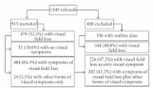
          Aims. To profile site of stroke/cerebrovascular accident, type and extent of field loss, treatment options, and outcome. Methods. Prospective multicentre cohort trial. Standardised referral and investigation protocol of visual parameters. Results. 915 patients were recruited with a mean age of 69 years (SD 14). 479 patients (52%) had visual field loss. 51 patients (10%) had no visual symptoms. Almost half of symptomatic patients ( n = 226) complained only of visual field loss: almost half ( n = 226) also had reading difficulty, blurred vision, diplopia, and perceptual difficulties. 31% ( n = 151) had visual field loss as their only visual impairment: 69% ( n = 328) had low vision, eye movement deficits, or visual perceptual difficulties. Occipital and parietal lobe strokes most commonly caused visual field loss. Treatment options included visual search training, visual awareness, typoscopes, substitutive prisms, low vision aids, refraction, and occlusive patches. At followup 15 patients (7.5%) had full recovery, 78 (39%) had improvement, and 104 (52%) had no recovery. Two patients (1%) had further decline of visual field. Patients with visual field loss had lower quality of life scores than stroke patients without visual impairment. Conclusions. Stroke survivors with visual field loss require assessment to accurately define type and extent of loss, diagnose coexistent visual impairments, and offer targeted treatment.

          Related collections

          Most cited references90

          • Record: found
          • Abstract: found
          • Article: not found

          Natural history of homonymous hemianopia.

          To describe the characteristics of spontaneous recovery of homonymous hemianopia (HH). The authors reviewed medical records of all patients with HH confirmed by formal visual field testing and seen in follow-up in their service between 1989 and 2004. Clinical characteristics, causes, neuroradiologic definition of lesion location, final outcome, and evolution of the visual field defects were recorded. The associations among final visual field defect outcome, time from injury, and clinical features were analyzed. A total of 254 patients with 263 HH were included in this study. Spontaneous visual field defect recovery was observed in 101 HH (38.4%). The likelihood of spontaneous recovery decreased with increasing time from injury to initial visual field testing (p = 0.0003). The probability of improvement was related to the time since injury (p = 0.0003) with a 50 to 60% chance of improvement for cases tested within 1 month after injury that decreased to about 20% for cases tested at 6 months after surgery. No other factor was found to correlate with the final outcome of the visual field defects. Improvement after 6 months from injury was mild and usually related to improvement of the underlying disease. Spontaneous improvement of homonymous hemianopia is seen in at least 50% of patients first seen within 1 month of injury. In most cases, the improvement occurs within the first 3 months from injury. Spontaneous improvement after 6 months postinjury should be interpreted with caution as it is most likely related to improvement of the underlying disease or to improvement in the patient's ability to perform visual field testing reliably.
            Bookmark
            • Record: found
            • Abstract: found
            • Article: not found

            Visual impairment and falls in older adults: the Blue Mountains Eye Study.

            To examine the association between visual impairment and falls in older people. Cross-sectional survey of eye disease with retrospective collection of falls data. Two postcode areas in the Blue Mountains west of Sydney, Australia. All people 49 years of age and older were invited to participate, 3654 (82.4%) of 4433 eligible residents took part, and 3299 answered questions about falls. Subjects had a detailed eye examination and answered questions about health and vision status, use of medication, and number of falls in the previous 12 months. Tests of visual function that had a statistically significant association with two or more falls after adjustment for confounders were visual acuity (prevalence ratio (PR) 1.9 for visual acuity worse than 20/30), contrast sensitivity (PR 1.2 for a 1-unit decrease at 6 cycles per degree), and suprathreshold visual field screening (PR 1.5 for 5 or more points missing). However, only visual acuity and contrast sensitivity were significantly associated with two or more falls per 1 standard deviation decrease. The presence of posterior subcapsular cataract (PR 2.1) and use of nonmiotic glaucoma medication (PR 2.0) had a statistically significant association with two or more falls; presence of age-related macular degeneration, diabetic retinopathy, and cortical or nuclear cataract did not. Visual impairment is strongly associated with two or more falls in older adults. In addition to poor visual acuity, visual factors such as reduced visual field, impaired contrast sensitivity, and the presence of cataract may explain this association.
              Bookmark
              • Record: found
              • Abstract: found
              • Article: not found

              Visual impairment following stroke: do stroke patients require vision assessment?

              the types of visual impairment followings stroke are wide ranging and encompass low vision, eye movement and visual field abnormalities, and visual perceptual difficulties. the purpose of this paper is to present a 1-year data set and identify the types of visual impairment occurring following stroke and their prevalence. a multi-centre prospective observation study was undertaken in 14 acute trust hospitals. Stroke survivors with a suspected visual difficulty were recruited. Standardised screening/referral and investigation forms were employed to document data on visual impairment specifically assessment of visual acuity, ocular pathology, eye alignment and movement, visual perception (including inattention) and visual field defects. three hundred and twenty-three patients were recruited with a mean age of 69 years [standard deviation (SD) 15]. Sixty-eight per cent had eye alignment/movement impairment, 49% had visual field impairment, 26.5% had low vision and 20.5% had perceptual difficulties. of patients referred with a suspected visual difficulty, only 8% had normal vision status confirmed on examination. Ninety-two per cent had visual impairment of some form confirmed which is considerably higher than previous publications and probably relates to the prospective, standardised investigation offered by specialist orthoptists. However, under-ascertainment of visual problems cannot be ruled out.
                Bookmark

                Author and article information

                Journal
                Biomed Res Int
                Biomed Res Int
                BMRI
                BioMed Research International
                Hindawi Publishing Corporation
                2314-6133
                2314-6141
                2013
                9 September 2013
                : 2013
                : 719096
                Affiliations
                1Department of Health Services Research, Whelan Building, University of Liverpool, Brownlow Hill, Liverpool L69 3GB, UK
                2Altnagelvin Hospitals HHS Trust, Altnagelvin BT47 6SB, UK
                3NHS Ayrshire and Arran, Ayr KA6 6DX, UK
                4Royal United Hospitals Bath NHS Trust, Bath BA1 3NG, UK
                5Fairfield Hospital, Bury BL9 7TD, UK
                6Durham and Darlington Hospitals NHS Foundation Trust, Durham DH1 5TW, UK
                7Ipswich Hospital NHS Trust, Ipswich IP4 5PD, UK
                8Gloucestershire Hospitals NHS Foundation Trust, Gloucester GL1 3NN, UK
                9St Helier General Hospital, Jersey JE1 3QS, UK
                10University Hospital NHS Trust, Nottingham BG7 2UH, UK
                11Oxford Radcliffe Hospitals NHS Trust, Oxford OX3 9DU, UK
                12Salford Royal NHS Foundation Trust, Salford M6 8HD, UK
                13Sheffield Teaching Hospitals NHS Foundation Trust, Sheffield S10 2JF, UK
                14Swindon and Marlborough NHS Trust, Swindon SN3 6BB, UK
                15Wrightington, Wigan and Leigh NHS Trust, Wigan WN1 2NN, UK
                16Worcestershire Acute Hospitals NHS Trust, Worcester WR5 1DD, UK
                Author notes

                Academic Editor: Alberto Raggi

                Author information
                http://orcid.org/0000-0002-0901-8413
                http://orcid.org/0000-0002-9426-1150
                http://orcid.org/0000-0002-2806-9144
                Article
                10.1155/2013/719096
                3782154
                24089687
                fcbfee46-b8dd-4502-a464-d3f0f6ff1ff4
                Copyright © 2013 Fiona J. Rowe et al.

                This is an open access article distributed under the Creative Commons Attribution License, which permits unrestricted use, distribution, and reproduction in any medium, provided the original work is properly cited.

                History
                : 23 April 2013
                : 23 June 2013
                : 22 July 2013
                Categories
                Research Article

                Comments

                Comment on this article