20
views
0
recommends
+1 Recommend
0 collections
    0
    shares
      • Record: found
      • Abstract: found
      • Article: found
      Is Open Access

      Long-term surgical outcomes of primary retropupillary iris claw intraocular lens implantation for the treatment of intraocular lens dislocation

      research-article

      Read this article at

      Bookmark
          There is no author summary for this article yet. Authors can add summaries to their articles on ScienceOpen to make them more accessible to a non-specialist audience.

          Abstract

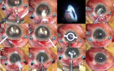
          We aimed to investigate the efficacy and safety of primary retropupillary iris claw intraocular lens (R-IOL) implantation in patients with complete intraocular lens (IOL) dislocation. In this single-center retrospective case series, we reviewed the medical records of patients who underwent R-IOL implantation surgery with pars plana vitrectomy for the treatment of IOL dislocation between September 2014 and July 2019. The primary outcome was change in visual acuity (VA) up to 24 months postoperatively. The secondary outcomes included changes in intraocular pressure (IOP), refractive errors, and endothelial cell count (ECC) over the same period. Data of 103 eyes (98 patients) were analyzed. The mean uncorrected VA was significantly improved at one month postoperatively (− 0.69 logMAR, P < 0.001), compared to the preoperative value. IOP (− 2.3 mmHg, P = 0.008) and ECC (− 333.4 cells/mm 2, P = 0.027) significantly decreased one month post-surgery and remained stable thereafter. Postoperative mean spherical equivalents were similar to the prediction error throughout the follow-up period. IOP elevation (n = 8, 7.8%), cystoid macular edema (n = 4, 3.9%), and dislocation of the R-IOL (n = 10, 9.7%) were managed successfully. Overall, primary R-IOL implantation with pars plana vitrectomy is effective and safe for correcting IOL dislocation due to various causes.

          Related collections

          Most cited references49

          • Record: found
          • Abstract: found
          • Article: not found

          Mechanisms of macular edema: Beyond the surface.

          Macular edema consists of intra- or subretinal fluid accumulation in the macular region. It occurs during the course of numerous retinal disorders and can cause severe impairment of central vision. Major causes of macular edema include diabetes, branch and central retinal vein occlusion, choroidal neovascularization, posterior uveitis, postoperative inflammation and central serous chorioretinopathy. The healthy retina is maintained in a relatively dehydrated, transparent state compatible with optimal light transmission by multiple active and passive systems. Fluid accumulation results from an imbalance between processes governing fluid entry and exit, and is driven by Starling equation when inner or outer blood-retinal barriers are disrupted. The multiple and intricate mechanisms involved in retinal hydro-ionic homeostasis, their molecular and cellular basis, and how their deregulation lead to retinal edema, are addressed in this review. Analyzing the distribution of junction proteins and water channels in the human macula, several hypotheses are raised to explain why edema forms specifically in the macular region. "Pure" clinical phenotypes of macular edema, that result presumably from a single causative mechanism, are detailed. Finally, diabetic macular edema is investigated, as a complex multifactorial pathogenic example. This comprehensive review on the current understanding of macular edema and its mechanisms opens perspectives to identify new preventive and therapeutic strategies for this sight-threatening condition.
            Bookmark
            • Record: found
            • Abstract: found
            • Article: not found

            Long-term outcome of combined pars plana vitrectomy and scleral fixated sutured posterior chamber intraocular lens implantation.

            To investigate the long-term visual outcome and the complication rate following transscleral suture fixation of posterior chamber intraocular lenses (sutured PC-IOLs). A retrospective case-series descriptive study. Records of patients who underwent combined pars plana vitrectomy and sutured PC-IOLs at Moorfields Eye Hospital and who had at least 12 months of follow-up were examined for recorded complications. Sixty-one eyes of 48 patients (33 males and 15 females) were identified and included in the analysis, with mean follow-up of 6 years. The mean final best-corrected visual acuity remained at preoperative levels (P=.211) and was largely determined by the underlying ocular pathology before sutured PC-IOL. Overall 30 of 61 (49%) eyes, two or more procedures were performed to reverse a significant peri- or postoperative complication. Breakage of polypropylene sutures was the main indication accounting for 17 of 30 (57%) of those reoperations. Subgroup analysis showed that younger patients were more likely to suffer the above complication (P=.009). The multivariate analysis also showed that longer follow-up was significantly associated with suture breakage (P=.014), with the mean time to breakage approximately 4 years after surgery. Long-term follow-up of patients undergoing sutured PC-IOLs appears to be associated with a high rate of postoperative complications and significant need for further surgery, which should be discussed during their informed consent process.
              Bookmark
              • Record: found
              • Abstract: found
              • Article: not found

              Clinical pseudophakic cystoid macular edema. Risk factors for development and duration after treatment.

              To characterize the incidence, duration, and risk factors for and outcome of cystoid macular edema (CME) after cataract surgery and investigate the effects of treatment regimens on visual outcome and duration. University-based comprehensive ophthalmology practice. This study included 1659 consecutive cataract surgeries performed by residents between 2001 and 2006. Cases were classified according to the presence of CME. Subset analysis excluded patients with diabetes mellitus (DM). The CME groups were analyzed according to type of treatment to compare duration of CME and final best corrected visual acuity. The incidence of postoperative CME was 2.35% (39/1659), and history of retinal vein occlusion (RVO) was predictive of postoperative CME (odds ratio [OR], 47.12; P<.001). When patients with DM were excluded, the incidence of CME was 2.14% (29/1357) and history of RVO (OR, 31.75; P<.001), epiretinal membrane (ERM) (OR, 4.93; P<.03), and preoperative prostaglandin use (OR, 12.45; P<.04) were predictive of postoperative CME. Patients with DM and/or intraoperative complications did not have an increased risk for CME when treated with prophylactic postoperative nonsteroidal antiinflammatory drugs (NSAIDs) for 3 months. Groups treated with NSAIDs plus a steroid had significantly shorter resolution times than the untreated group (P = .004). A history of RVO, ERM, and preoperative prostaglandin use were associated with an increased risk for pseudophakic CME. Treatment with NSAIDs plus steroids was associated with faster resolution of CME than no treatment. Treating high-risk patients with NSAIDs after cataract surgery decreases the incidence of postoperative CME to that of patients who are not at high risk.
                Bookmark

                Author and article information

                Contributors
                minkim76@gmail.com
                Journal
                Sci Rep
                Sci Rep
                Scientific Reports
                Nature Publishing Group UK (London )
                2045-2322
                12 January 2021
                12 January 2021
                2021
                : 11
                : 726
                Affiliations
                [1 ]GRID grid.15444.30, ISNI 0000 0004 0470 5454, Department of Ophthalmology, Institute of Vision Research, Severance Eye Hospital, , Yonsei University College of Medicine, ; 50-1, Yonseiro, Seodaemun-gu, Seoul, 03722 Korea
                [2 ]GRID grid.15444.30, ISNI 0000 0004 0470 5454, Department of Ophthalmology, Institute of Vision Research, Gangnam Severance Hospital, , Yonsei University College of Medicine, ; 211, Eonjuro, Gangnam-gu, Seoul, 06273 Korea
                Article
                80292
                10.1038/s41598-020-80292-3
                7804091
                33436860
                fcc7ea5c-461a-4b0e-8a35-104689a99e2e
                © The Author(s) 2021

                Open Access This article is licensed under a Creative Commons Attribution 4.0 International License, which permits use, sharing, adaptation, distribution and reproduction in any medium or format, as long as you give appropriate credit to the original author(s) and the source, provide a link to the Creative Commons licence, and indicate if changes were made. The images or other third party material in this article are included in the article's Creative Commons licence, unless indicated otherwise in a credit line to the material. If material is not included in the article's Creative Commons licence and your intended use is not permitted by statutory regulation or exceeds the permitted use, you will need to obtain permission directly from the copyright holder. To view a copy of this licence, visit http://creativecommons.org/licenses/by/4.0/.

                History
                : 5 August 2020
                : 9 November 2020
                Funding
                Funded by: FundRef http://dx.doi.org/10.13039/501100008005, Yonsei University College of Medicine;
                Award ID: 2017-32-0037
                Award Recipient :
                Categories
                Article
                Custom metadata
                © The Author(s) 2021

                Uncategorized
                lens diseases,outcomes research
                Uncategorized
                lens diseases, outcomes research

                Comments

                Comment on this article