13
views
0
recommends
+1 Recommend
3 collections
    0
    shares
      • Record: found
      • Abstract: found
      • Article: found

      Morphea induced by SARS‐CoV‐2 infection: A case report

      letter

      Read this article at

      ScienceOpenPublisherPMC
      Bookmark
          There is no author summary for this article yet. Authors can add summaries to their articles on ScienceOpen to make them more accessible to a non-specialist audience.

          Abstract

          Dear Editor, The widespread diffusion of severe acute respiratory syndrome coronavirus‐2 (SARS‐CoV‐2), a member of the coronavirus family responsible for Coronavirus disease (COVID‐19), is still in the headlines for the impressive health and economic burden worldwide. The immune response against this novel virus is still undergoing extensive research, and the possible long‐term outcomes remain basically unknown. Immunity plays a key role in the defense against several microorganisms, and SARS‐CoV‐2 is no different. The infections can result in a hyperactive immune response with excessive inflammatory reactions. 1 These are generally accompanied by the release of impressive amounts of proinflammatory cytokines, an event known as “cytokine storm,” that is correlated with lung damage, organ failure, and a negative prognosis. 2 , 3 The possibility that these immunologic derangements could result in the induction and/or the exacerbation of other immune‐mediated diseases, including cutaneous ones, has not been completely investigated. We report the case of a 61‐year‐old female patient referred to our dermatologic department for the development of asymptomatic bilateral sclerotic cutaneous lesions on the forearms which appeared 2 months before. The physical examination revealed the presence of several brownish and violaceous plaques with mild erythematous borders on forearms, with sclerotic appearance, partially tending to confluence. The lesions showed a symmetrical 
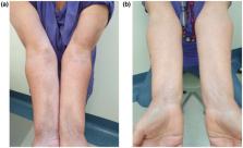
and bilateral distribution and were about 5–10 cm in diameter (Fig. 1a). The patient referred that the skin manifestations started about 1 month after the dismissal from hospitalization for COVID‐19 pneumonia (confirmed through RT‐PCR). Her familiar and personal history was negative for autoimmune and chronic inflammatory skin disorders. On suspicion of localized morphea, blood examination tests and skin biopsy were performed. Blood tests, including antinuclear antibodies (ANA), antibodies to single‐stranded DNA (a‐ssDNA), autoantibodies to extractable nuclear antigens (ENAs), and COVID‐19 swab test, were negative excluding systemic involvement. Histological examination revealed the presence of thin epidermis with moderate dermal sclerosis and thickening of collagen fibers, confirming the diagnosis of morphea. Figure 1 Clinical manifestations: (a) the presence of several brownish and violaceous plaques with mild erythematous borders on forearms, with sclerotic appearance, partially tending to confluence. (b) Complete clinical resolution after 16 weeks of treatment with clobetasol ointment and vitamin E emollient A topical therapy with a high‐potency steroid (clobetasol ointment) and vitamin E emollient was started, with a remarkable improvement after 8 weeks and a complete clinical resolution after 16 weeks of treatment (Fig. 1b). Morphea, also known as localized scleroderma, is an inflammatory skin condition that is characterized by the presentation of single or multiple inflammatory or sclerotic plaques. 4 While the pathogenesis remains largely unknown, several factors can contribute to the emergence of autoimmune disease including the genetic predisposition, environmental triggers such as bacterial and viral infections, and intrinsic factors, such as hormonal and immunologic dysregulation. 5 In this case, the negative personal anamnesis for autoimmune disorders and the development of cutaneous manifestations after COVID‐19 infection suggested a possible link between autoimmune disease and SARS‐CoV‐2. Some authors proposed that SARS‐CoV‐2 could act as a trigger in the development of organ‐specific autoimmune disorders, in genetic predisposed subjects. 6 In particular, a molecular mimicry phenomenon between virus and human protein has been hypothesized. Hence, the exaggerated activation of immune system against the virus could induce a cross‐reaction with auto‐antigens in common with viral peptides, leading to an autoimmune dysfunction. 7 Recently, some authors reported the development and/or worsening of inflammatory skin diseases after COVID‐19 infection, such as atopic dermatitis 8 and psoriasis, 9 induced by abnormal activation of the immune system in response to virus and consequent inflammatory pathways. To the best of our knowledge, in the literature, there are no reports of morphea triggered by COVID‐19 infection. We report this case to underline the importance of this clinical condition during the COVID‐19 pandemic for the dermatologists, in order to provide a proper diagnosis and a correct therapeutic management.

          Related collections

          Most cited references10

          • Record: found
          • Abstract: found
          • Article: not found

          Clinical features of patients infected with 2019 novel coronavirus in Wuhan, China

          Summary Background A recent cluster of pneumonia cases in Wuhan, China, was caused by a novel betacoronavirus, the 2019 novel coronavirus (2019-nCoV). We report the epidemiological, clinical, laboratory, and radiological characteristics and treatment and clinical outcomes of these patients. Methods All patients with suspected 2019-nCoV were admitted to a designated hospital in Wuhan. We prospectively collected and analysed data on patients with laboratory-confirmed 2019-nCoV infection by real-time RT-PCR and next-generation sequencing. Data were obtained with standardised data collection forms shared by WHO and the International Severe Acute Respiratory and Emerging Infection Consortium from electronic medical records. Researchers also directly communicated with patients or their families to ascertain epidemiological and symptom data. Outcomes were also compared between patients who had been admitted to the intensive care unit (ICU) and those who had not. Findings By Jan 2, 2020, 41 admitted hospital patients had been identified as having laboratory-confirmed 2019-nCoV infection. Most of the infected patients were men (30 [73%] of 41); less than half had underlying diseases (13 [32%]), including diabetes (eight [20%]), hypertension (six [15%]), and cardiovascular disease (six [15%]). Median age was 49·0 years (IQR 41·0–58·0). 27 (66%) of 41 patients had been exposed to Huanan seafood market. One family cluster was found. Common symptoms at onset of illness were fever (40 [98%] of 41 patients), cough (31 [76%]), and myalgia or fatigue (18 [44%]); less common symptoms were sputum production (11 [28%] of 39), headache (three [8%] of 38), haemoptysis (two [5%] of 39), and diarrhoea (one [3%] of 38). Dyspnoea developed in 22 (55%) of 40 patients (median time from illness onset to dyspnoea 8·0 days [IQR 5·0–13·0]). 26 (63%) of 41 patients had lymphopenia. All 41 patients had pneumonia with abnormal findings on chest CT. Complications included acute respiratory distress syndrome (12 [29%]), RNAaemia (six [15%]), acute cardiac injury (five [12%]) and secondary infection (four [10%]). 13 (32%) patients were admitted to an ICU and six (15%) died. Compared with non-ICU patients, ICU patients had higher plasma levels of IL2, IL7, IL10, GSCF, IP10, MCP1, MIP1A, and TNFα. Interpretation The 2019-nCoV infection caused clusters of severe respiratory illness similar to severe acute respiratory syndrome coronavirus and was associated with ICU admission and high mortality. Major gaps in our knowledge of the origin, epidemiology, duration of human transmission, and clinical spectrum of disease need fulfilment by future studies. Funding Ministry of Science and Technology, Chinese Academy of Medical Sciences, National Natural Science Foundation of China, and Beijing Municipal Science and Technology Commission.
            Bookmark
            • Record: found
            • Abstract: found
            • Article: not found

            Clinical predictors of mortality due to COVID-19 based on an analysis of data of 150 patients from Wuhan, China

            Dear Editor, The rapid emergence of COVID-19 in Wuhan city, Hubei Province, China, has resulted in thousands of deaths [1]. Many infected patients, however, presented mild flu-like symptoms and quickly recover [2]. To effectively prioritize resources for patients with the highest risk, we identified clinical predictors of mild and severe patient outcomes. Using the database of Jin Yin-tan Hospital and Tongji Hospital, we conducted a retrospective multicenter study of 68 death cases (68/150, 45%) and 82 discharged cases (82/150, 55%) with laboratory-confirmed infection of SARS-CoV-2. Patients met the discharge criteria if they had no fever for at least 3 days, significantly improved respiratory function, and had negative SARS-CoV-2 laboratory test results twice in succession. Case data included demographics, clinical characteristics, laboratory results, treatment options and outcomes. For statistical analysis, we represented continuous measurements as means (SDs) or as medians (IQRs) which compared with Student’s t test or the Mann–Whitney–Wilcoxon test. Categorical variables were expressed as numbers (%) and compared by the χ 2 test or Fisher’s exact test. The distribution of the enrolled patients’ age is shown in Fig. 1a. There was a significant difference in age between the death group and the discharge group (p < 0.001) but no difference in the sex ratio (p = 0.43). A total of 63% (43/68) of patients in the death group and 41% (34/82) in the discharge group had underlying diseases (p = 0.0069). It should be noted that patients with cardiovascular diseases have a significantly increased risk of death when they are infected with SARS-CoV-2 (p < 0.001). A total of 16% (11/68) of the patients in the death group had secondary infections, and 1% (1/82) of the patients in the discharge group had secondary infections (p = 0.0018). Laboratory results showed that there were significant differences in white blood cell counts, absolute values of lymphocytes, platelets, albumin, total bilirubin, blood urea nitrogen, blood creatinine, myoglobin, cardiac troponin, C-reactive protein (CRP) and interleukin-6 (IL-6) between the two groups (Fig. 1b and Supplementary Table 1). Fig. 1 a Age distribution of patients with confirmed COVID-19; b key laboratory parameters for the outcomes of patients with confirmed COVID-19; c interval from onset of symptom to death of patients with confirmed COVID-19; d summary of the cause of death of 68 died patients with confirmed COVID-19 The survival times of the enrolled patients in the death group were analyzed. The distribution of survival time from disease onset to death showed two peaks, with the first one at approximately 14 days (22 cases) and the second one at approximately 22 days (17 cases) (Fig. 1c). An analysis of the cause of death was performed. Among the 68 fatal cases, 36 patients (53%) died of respiratory failure, five patients (7%) with myocardial damage died of circulatory failure, 22 patients (33%) died of both, and five remaining died of an unknown cause (Fig. 1d). Based on the analysis of the clinical data, we confirmed that some patients died of fulminant myocarditis. In this study, we first reported that the infection of SARS-CoV-2 may cause fulminant myocarditis. Given that fulminant myocarditis is characterized by a rapid progress and a severe state of illness [3], our results should alert physicians to pay attention not only to the symptoms of respiratory dysfunction but also the symptoms of cardiac injury. Further, large-scale studies and the studies on autopsy are needed to confirm our analysis. In conclusion, predictors of a fatal outcome in COVID-19 cases included age, the presence of underlying diseases, the presence of secondary infection and elevated inflammatory indicators in the blood. The results obtained from this study also suggest that COVID-19 mortality might be due to virus-activated “cytokine storm syndrome” or fulminant myocarditis. Electronic supplementary material Below is the link to the electronic supplementary material. Supplementary material 1 (DOCX 38 kb)
              Bookmark
              • Record: found
              • Abstract: found
              • Article: found
              Is Open Access

              The COVID-19 Cytokine Storm; What We Know So Far

              COVID-19 is a rapidly spreading global threat that has been declared as a pandemic by the WHO. COVID-19 is transmitted via droplets or direct contact and infects the respiratory tract resulting in pneumonia in most of the cases and acute respiratory distress syndrome (ARDS) in about 15 % of the cases. Mortality in COVID-19 patients has been linked to the presence of the so-called “cytokine storm” induced by the virus. Excessive production of proinflammatory cytokines leads to ARDS aggravation and widespread tissue damage resulting in multi-organ failure and death. Targeting cytokines during the management of COVID-19 patients could improve survival rates and reduce mortality.
                Bookmark

                Author and article information

                Contributors
                maria.mariano@ifo.gov.it
                Journal
                Int J Dermatol
                Int J Dermatol
                10.1111/(ISSN)1365-4632
                IJD
                International Journal of Dermatology
                John Wiley and Sons Inc. (Hoboken )
                0011-9059
                1365-4632
                15 November 2021
                15 November 2021
                : 10.1111/ijd.15983
                Affiliations
                [ 1 ] San Gallicano Dermatological Institute – IRCCS Rome Italy
                Author information
                https://orcid.org/0000-0002-7964-3388
                Article
                IJD15983
                10.1111/ijd.15983
                8653008
                34783020
                fea59559-a519-47a8-8d7f-d5534de05cf9
                © 2021 the International Society of Dermatology

                This article is being made freely available through PubMed Central as part of the COVID-19 public health emergency response. It can be used for unrestricted research re-use and analysis in any form or by any means with acknowledgement of the original source, for the duration of the public health emergency.

                History
                : 05 October 2021
                : 11 August 2021
                : 28 October 2021
                Page count
                Figures: 1, Tables: 0, Pages: 2, Words: 911
                Categories
                Correspondence
                Correspondence
                Custom metadata
                2.0
                corrected-proof
                Converter:WILEY_ML3GV2_TO_JATSPMC version:6.0.9 mode:remove_FC converted:08.12.2021

                Comments

                Comment on this article