19
views
0
recommends
+1 Recommend
0 collections
    0
    shares
      • Record: found
      • Abstract: found
      • Article: not found

      Cardiovascular Determinants of Aerobic Exercise Capacity in Adults With Type 2 Diabetes

      research-article

      Read this article at

      ScienceOpenPublisherPMC
      Bookmark
          There is no author summary for this article yet. Authors can add summaries to their articles on ScienceOpen to make them more accessible to a non-specialist audience.

          Abstract

          OBJECTIVE

          To assess the relationship between subclinical cardiac dysfunction and aerobic exercise capacity (peak VO 2) in adults with type 2 diabetes (T2D), a group at high risk of developing heart failure.

          RESEARCH DESIGN AND METHODS

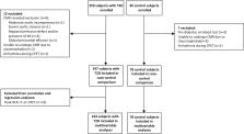
          Cross-sectional study. We prospectively enrolled a multiethnic cohort of asymptomatic adults with T2D and no history, signs, or symptoms of cardiovascular disease. Age-, sex-, and ethnicity-matched control subjects were recruited for comparison. Participants underwent bioanthropometric profiling, cardiopulmonary exercise testing, and cardiovascular magnetic resonance with adenosine stress perfusion imaging. Multivariable linear regression analysis was undertaken to identify independent associations between measures of cardiovascular structure and function and peak VO 2.

          RESULTS

          A total of 247 adults with T2D (aged 51.8 ± 11.9 years, 55% males, 37% black or south Asian ethnicity, HbA 1c 7.4 ± 1.1% [57 ± 12 mmol/mol], and duration of diabetes 61 [32–120] months) and 78 control subjects were included. Subjects with T2D had increased concentric left ventricular remodeling, reduced myocardial perfusion reserve (MPR), and markedly lower aerobic exercise capacity (peak VO 2 18.0 ± 6.6 vs. 27.8 ± 9.0 mL/kg/min; P < 0.001) compared with control subjects. In a multivariable linear regression model containing age, sex, ethnicity, smoking status, and systolic blood pressure, only MPR (β = 0.822; P = 0.006) and left ventricular diastolic filling pressure (E/e′) (β = −0.388; P = 0.001) were independently associated with peak VO 2 in subjects with T2D.

          CONCLUSIONS

          In a multiethnic cohort of asymptomatic people with T2D, MPR and diastolic function are key determinants of aerobic exercise capacity, independent of age, sex, ethnicity, smoking status, or blood pressure.

          Related collections

          Most cited references37

          • Record: found
          • Abstract: found
          • Article: not found

          Exercise hemodynamics enhance diagnosis of early heart failure with preserved ejection fraction.

          When advanced, heart failure with preserved ejection fraction (HFpEF) is readily apparent. However, diagnosis of earlier disease may be challenging because exertional dyspnea is not specific for heart failure, and biomarkers and hemodynamic indicators of volume overload may be absent at rest. Patients with exertional dyspnea and ejection fraction >50% were referred for hemodynamic catheterization. Those with no significant coronary disease, normal brain natriuretic peptide assay, and normal resting hemodynamics (mean pulmonary artery pressure <25 mm Hg and pulmonary capillary wedge pressure [PCWP] <15 mm Hg) (n=55) underwent exercise study. The exercise PCWP was used to classify patients as having HFpEF (PCWP ≥25 mm Hg) (n=32) or noncardiac dyspnea (PCWP <25 mm Hg) (n=23). At rest, patients with HFpEF had higher resting pulmonary artery pressure and PCWP, although all values fell within normal limits. Exercise-induced elevation in PCWP in HFpEF was confirmed by greater increases in left ventricular end-diastolic pressure and was associated with blunted increases in heart rate, systemic vasodilation, and cardiac output. Exercise-induced pulmonary hypertension was present in 88% of patients with HFpEF and was related principally to elevated PCWP, as pulmonary vascular resistances dropped similarly in both groups. Exercise PCWP and pulmonary artery systolic pressure were highly correlated. An exercise pulmonary artery systolic pressure ≥45 mm Hg identified HFpEF with 96% sensitivity and 95% specificity. Euvolemic patients with exertional dyspnea, normal brain natriuretic peptide, and normal cardiac filling pressures at rest may have markedly abnormal hemodynamic responses during exercise, suggesting that chronic symptoms are related to heart failure. Earlier and more accurate diagnosis using exercise hemodynamics may allow better targeting of interventions to treat and prevent HFpEF progression.
            Bookmark
            • Record: found
            • Abstract: found
            • Article: not found

            Association between coronary vascular dysfunction and cardiac mortality in patients with and without diabetes mellitus.

            Diabetes mellitus increases the risk of adverse cardiac outcomes and is considered a coronary artery disease (CAD) equivalent. We examined whether coronary vascular dysfunction, an early manifestation of CAD, accounts for increased risk among diabetics compared with nondiabetics. A total of 2783 consecutive patients (1172 diabetics and 1611 nondiabetics) underwent quantification of coronary flow reserve (CFR; CFR=stress divided by rest myocardial blood flow) by positron emission tomography and were followed up for a median of 1.4 years (quartile 1-3, 0.7-3.2 years). The primary end point was cardiac death. Impaired CFR (below the median) was associated with an adjusted 3.2- and 4.9-fold increase in the rate of cardiac death for diabetics and nondiabetics, respectively (P=0.0004). Addition of CFR to clinical and imaging risk models improved risk discrimination for both diabetics and nondiabetics (c index, 0.77-0.79, P=0.04; 0.82-0.85, P=0.03, respectively). Diabetic patients without known CAD with impaired CFR experienced a rate of cardiac death comparable to that for nondiabetic patients with known CAD (2.8%/y versus 2.0%/y; P=0.33). Conversely, diabetics without known CAD and preserved CFR had very low annualized cardiac mortality, which was similar to patients without known CAD or diabetes mellitus and normal stress perfusion and systolic function (0.3%/y versus 0.5%/y; P=0.65). Coronary vasodilator dysfunction is a powerful, independent correlate of cardiac mortality among both diabetics and nondiabetics and provides meaningful incremental risk stratification. Among diabetic patients without CAD, those with impaired CFR have event rates comparable to those of patients with prior CAD, whereas those with preserved CFR have event rates comparable to those of nondiabetics.
              Bookmark
              • Record: found
              • Abstract: found
              • Article: not found

              Prevalence of ventricular diastolic dysfunction in asymptomatic, normotensive patients with diabetes mellitus.

              To determine the prevalence of left ventricular diastolic dysfunction in asymptomatic, normotensive patients with type 2 diabetes mellitus, we studied 61 consecutive normotensive patients with type 2 diabetes using conventional Doppler echocardiography at rest (deceleration time, isovolumic relaxation time, early diastolic velocity [E]/peak atrial systolic velocity [A] ratio), and during the Valsalva maneuver. In addition, mitral annular velocity and velocity of flow propagation were assessed in all patients using tissue Doppler imaging (TDI) and color M-mode echocardiography. A standard resting echocardiogram excluded significant valvular disease and stress echocardiography excluded significant coronary artery disease in those with diastolic dysfunction. Diastolic dysfunction was found in 43 of 57 patients (75%) when all of the above echocardiographic techniques were used. TDI detected diastolic dysfunction more often (63%) than any other echocardiographic approach. Thus, the prevalence of left ventricular diastolic dysfunction in asymptomatic, normotensive patients with type 2 diabetes without significant coronary artery disease is much higher than previously suspected. TDI markedly improved the echocardiographic detection of diastolic dysfunction in asymptomatic patients with type 2 diabetes.
                Bookmark

                Author and article information

                Journal
                Diabetes Care
                Diabetes Care
                diacare
                dcare
                Diabetes Care
                Diabetes Care
                American Diabetes Association
                0149-5992
                1935-5548
                September 2020
                17 July 2020
                17 July 2020
                : 43
                : 9
                : 2248-2256
                Affiliations
                [1] 1Department of Cardiovascular Sciences, University of Leicester and the NIHR Leicester Biomedical Research Centre, Leicester, U.K.
                [2] 2Diabetes Research Centre, University of Leicester and the NIHR Leicester Biomedical Research Centre, Leicester, U.K.
                [3] 3Diabetes Department, Royal Derby Hospital, University Hospitals of Derby and Burton NHS Foundation Trust, Derby, U.K.
                [4] 4NIHR Leeds Biomedical Research Centre, Leeds, U.K.
                [5] 5National Heart, Lung, and Blood Institute, Bethesda, MD
                Author notes
                Corresponding author: Gerry P. McCann, gpm12@ 123456leicester.ac.uk
                Author information
                https://orcid.org/0000-0002-1740-9270
                https://orcid.org/0000-0003-0395-7329
                https://orcid.org/0000-0003-2032-8938
                https://orcid.org/0000-0002-3932-3339
                https://orcid.org/0000-0003-2343-7099
                Article
                200706
                10.2337/dc20-0706
                7440912
                32680830
                ff9319b5-ba6b-4b6d-9c3c-07195b80a972
                © 2020 by the American Diabetes Association

                Readers may use this article as long as the work is properly cited, the use is educational and not for profit, and the work is not altered. More information is available at https://www.diabetesjournals.org/content/license.

                History
                : 1 April 2020
                : 9 June 2020
                Page count
                Figures: 2, Tables: 2, Equations: 0, References: 46, Pages: 9
                Funding
                Funded by: NIHR Research Trainees Coordinating Centre, DOI https://dx.doi.org/10.13039/501100000659;
                Award ID: CDF 2014-07-045
                Funded by: British Heart Foundation, DOI https://dx.doi.org/10.13039/501100000274;
                Award ID: FS/16/47/32190
                Funded by: Medical Research Council, DOI https://dx.doi.org/10.13039/501100000265;
                Award ID: Interdisciplinary Bridging Award
                Funded by: Novo Nordisk, DOI https://dx.doi.org/10.13039/501100004191;
                Categories
                Cardiovascular and Metabolic Risk

                Endocrinology & Diabetes
                Endocrinology & Diabetes

                Comments

                Comment on this article