12
views
0
recommends
+1 Recommend
0 collections
    0
    shares
      • Record: found
      • Abstract: found
      • Article: found
      Is Open Access

      Three-dimensional bioprinting of multicell-laden scaffolds containing bone morphogenic protein-4 for promoting M2 macrophage polarization and accelerating bone defect repair in diabetes mellitus

      research-article

      Read this article at

      Bookmark
          There is no author summary for this article yet. Authors can add summaries to their articles on ScienceOpen to make them more accessible to a non-specialist audience.

          Abstract

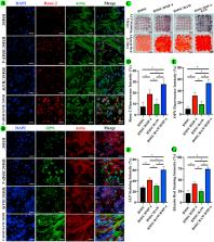
          Critical-sized bone defect repair in patients with diabetes mellitus remains a challenge in clinical treatment because of dysfunction of macrophage polarization and the inflammatory microenvironment in the bone defect region. Three-dimensional (3D) bioprinted scaffolds loaded with live cells and bioactive factors can improve cell viability and the inflammatory microenvironment and further accelerating bone repair. Here, we used modified bioinks comprising gelatin, gelatin methacryloyl (GelMA), and 4-arm poly (ethylene glycol) acrylate (PEG) to fabricate 3D bioprinted scaffolds containing BMSCs, RAW264.7 macrophages, and BMP-4-loaded mesoporous silica nanoparticles (MSNs). Addition of MSNs effectively improved the mechanical strength of GelMA/gelatin/PEG scaffolds. Moreover, MSNs sustainably released BMP-4 for long-term effectiveness. In 3D bioprinted scaffolds, BMP-4 promoted the polarization of RAW264.7 to M2 macrophages, which secrete anti-inflammatory factors and thereby reduce the levels of pro-inflammatory factors. BMP-4 released from MSNs and BMP-2 secreted from M2 macrophages collectively stimulated the osteogenic differentiation of BMSCs in the 3D bioprinted scaffolds. Furthermore, in calvarial critical-size defect models of diabetic rats, 3D bioprinted scaffolds loaded with MSNs/BMP-4 induced M2 macrophage polarization and improved the inflammatory microenvironment. And 3D bioprinted scaffolds with MSNs/BMP-4, BMSCs, and RAW264.7 cells significantly accelerated bone repair. In conclusion, our results indicated that implanting 3D bioprinted scaffolds containing MSNs/BMP-4, BMSCs, and RAW264.7 cells in bone defects may be an effective method for improving diabetic bone repair, owing to the direct effects of BMP-4 on promoting osteogenesis of BMSCs and regulating M2 type macrophage polarization to improve the inflammatory microenvironment and secrete BMP-2.

          Graphical abstract

          Highlights

          • The GelMA/gelatin/PEG/MSN composite bioinks showed satisfactory printability, mechanical stability, and biocompatibility.

          • The sustained release of BMP-4 from MSNs induced M2 macrophage polarization and thereby inhibited inflammatory reactions.

          • Loading of BMP-4 and secretion of BMP-2 by M2 type macrophages accelerated bone repair in DM bone defects.

          Related collections

          Most cited references46

          • Record: found
          • Abstract: found
          • Article: not found

          Bioink properties before, during and after 3D bioprinting.

          Bioprinting is a process based on additive manufacturing from materials containing living cells. These materials, often referred to as bioink, are based on cytocompatible hydrogel precursor formulations, which gel in a manner compatible with different bioprinting approaches. The bioink properties before, during and after gelation are essential for its printability, comprising such features as achievable structural resolution, shape fidelity and cell survival. However, it is the final properties of the matured bioprinted tissue construct that are crucial for the end application. During tissue formation these properties are influenced by the amount of cells present in the construct, their proliferation, migration and interaction with the material. A calibrated computational framework is able to predict the tissue development and maturation and to optimize the bioprinting input parameters such as the starting material, the initial cell loading and the construct geometry. In this contribution relevant bioink properties are reviewed and discussed on the example of most popular bioprinting approaches. The effect of cells on hydrogel processing and vice versa is highlighted. Furthermore, numerical approaches were reviewed and implemented for depicting the cellular mechanics within the hydrogel as well as for prediction of mechanical properties to achieve the desired hydrogel construct considering cell density, distribution and material-cell interaction.
            Bookmark
            • Record: found
            • Abstract: found
            • Article: not found

            A simple and high-resolution stereolithography-based 3D bioprinting system using visible light crosslinkable bioinks.

            Bioprinting is a rapidly developing technique for biofabrication. Because of its high resolution and the ability to print living cells, bioprinting has been widely used in artificial tissue and organ generation as well as microscale living cell deposition. In this paper, we present a low-cost stereolithography-based bioprinting system that uses visible light crosslinkable bioinks. This low-cost stereolithography system was built around a commercial projector with a simple water filter to prevent harmful infrared radiation from the projector. The visible light crosslinking was achieved by using a mixture of polyethylene glycol diacrylate (PEGDA) and gelatin methacrylate (GelMA) hydrogel with eosin Y based photoinitiator. Three different concentrations of hydrogel mixtures (10% PEG, 5% PEG + 5% GelMA, and 2.5% PEG + 7.5% GelMA, all w/v) were studied with the presented systems. The mechanical properties and microstructure of the developed bioink were measured and discussed in detail. Several cell-free hydrogel patterns were generated to demonstrate the resolution of the solution. Experimental results with NIH 3T3 fibroblast cells show that this system can produce a highly vertical 3D structure with 50 μm resolution and 85% cell viability for at least five days. The developed system provides a low-cost visible light stereolithography solution and has the potential to be widely used in tissue engineering and bioengineering for microscale cell patterning.
              Bookmark
              • Record: found
              • Abstract: not found
              • Article: not found

              Current State of Fabrication Technologies and Materials for Bone Tissue Engineering

                Bookmark

                Author and article information

                Contributors
                Journal
                Bioact Mater
                Bioactive Materials
                KeAi Publishing
                2452-199X
                25 September 2020
                March 2021
                25 September 2020
                : 6
                : 3
                : 757-769
                Affiliations
                [a ]Shanghai Key Laboratory of Orthopaedic Implants, Department of Orthopaedic Surgery, Shanghai Ninth People's Hospital, Shanghai JiaoTong University School of Medicine, No. 639 Zhizaoju Road, Shanghai, 200011, China
                [b ]Department of Radiology, Shanghai Ninth People's Hospital, Shanghai JiaoTong University School of Medicine, No. 639 Zhizaoju Road, Shanghai, 200011, China
                [c ]Department of Radiology, Minhang Hospital of Fudan University, Minhang Central Hospital, No. 170 Xinsong Road, Shanghai 201100, China
                [d ]Southwest JiaoTong University College of Medicine, No. 111 North 1st Section of Second Ring Road, Chengdu, 610031, China
                [e ]School of Biomedical Engineering, Shanghai JiaoTong University, No. 1956 Huashan Road, Shanghai, 200030, China
                [f ]Department of Sports Medicine, Shanghai Jiao Tong University Affiliated Sixth People's Hospital, No. 600, Yishan Road, Shanghai 200233, China
                [g ]Spine Institute, Shanghai University of Traditional Chinese Medicine, No.1200 Cailun Road, Shanghai 200032, China
                [h ]College of Chemistry, Chemical Engineering and Biotechnology, Donghua University, No. 2999, North Renmin Road, Shanghai 201620, China
                [i ]Department of Orthopaedics, Xinhua Hospital affiliated to Shanghai Jiaotong University School of Medicine, No.1665 Kongjiang Road, Shanghai, 200092, China
                Author notes
                []Corresponding author. wangjw@ 123456shsmu.edu.cn
                [∗∗ ]Corresponding author. litaoxyeyy@ 123456sina.com
                [∗∗∗ ]Corresponding author. zxj@ 123456dhu.edu.cn
                [1]

                Contributed equally to this work.

                Article
                S2452-199X(20)30203-6
                10.1016/j.bioactmat.2020.08.030
                7522044
                33024897
                01318fc2-cdff-4e89-a752-360506c28f96
                © 2020 The Authors. Publishing services by Elsevier B.V. on behalf of KeAi Communications Co., Ltd.

                This is an open access article under the CC BY-NC-ND license (http://creativecommons.org/licenses/by-nc-nd/4.0/).

                History
                : 15 June 2020
                : 9 August 2020
                : 23 August 2020
                Categories
                Article

                three-dimensional bioprinting,diabetic bone defect,bone morphogenic protein-4,macrophage polarization,bone regeneration

                Comments

                Comment on this article