21
views
0
recommends
+1 Recommend
1 collections
    0
    shares

      To submit to Bentham Journals, please click here

      • Record: found
      • Abstract: found
      • Article: found
      Is Open Access

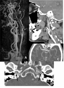
      Craniocervical Dissections: Radiologic Findings, Pitfalls, Mimicking Diseases: A Pictorial Review

      review-article

      Read this article at

      Bookmark
          There is no author summary for this article yet. Authors can add summaries to their articles on ScienceOpen to make them more accessible to a non-specialist audience.

          Abstract

          Background:

          Craniocervical Dissections (CCD) are a crucial emergency state causing 20% of strokes in patients under the age of 45. Although DSA (digital substraction angiography) is regarded as the gold standard, noninvasive methods of CT, CTA and MRI, MRA are widely used for diagnosis.

          Aim:

          Our aim is to illustrate noninvasive imaging findings in CCD.

          Conclusion:

          Emphasizing on diagnostic pitfalls, limitations and mimicking diseases.

          Related collections

          Most cited references43

          • Record: found
          • Abstract: found
          • Article: not found

          Blunt carotid arterial injuries: implications of a new grading scale.

          Blunt carotid arterial injuries (BCI) have the potential for devastating outcomes. A paucity of literature and the absence of a formal BCI grading scale have been major impediments to the formulation of sound practice guidelines. We reviewed our experience with 109 BCI and developed a grading scale with prognostic and therapeutic implications. Patients admitted to a Level I trauma center were evaluated with cerebral arteriography if they exhibited signs or symptoms of BCI or met criteria for screening. Patients with BCI were treated with heparin unless they had contraindications, and follow-up arteriography was performed at 7 to 10 days. Endovascular stents were deployed selectively. A prospective database was used to track the patients. A total of 76 patients were diagnosed with 109 BCI. Two-thirds of mild intimal injuries (grade I) healed, regardless of therapy. Dissections or hematomas with luminal stenosis (grade II) progressed, despite heparin therapy in 70% of cases. Only 8% of pseudoaneurysms (grade III) healed with heparin, but 89% resolved after endovascular stent placement. Occlusions (grade IV) did not recanalize in the early postinjury period. Grade V injuries (transections) were lethal and refractory to intervention. Stroke risk increased with injury grade. Severe head injuries (Glasgow Coma Scale score < or =6) were found in 46% of patients and confounded evaluation of neurologic outcomes. This BCI grading scale has prognostic and therapeutic implications. Nonoperative treatment options for grade I BCI should be evaluated in prospective, randomized trials. Accessible grade II, III, IV, and V lesions should be surgically repaired. Inaccessible grade II, III, and IV injuries should be treated with systemic anticoagulation. Endovascular techniques may be the only recourse in high grade V injuries and warrant controlled evaluation in the treatment of grade III BCI.
            Bookmark
            • Record: found
            • Abstract: found
            • Article: not found

            Incidence and outcome of cervical artery dissection: a population-based study.

            Incidence rates for internal carotid artery dissection (ICAD) have been reported to be 2.6 to 2.9 per 100,000, but reliable epidemiologic data for vertebral artery dissection (VAD) are not available. To determine the incidence rate of cervical artery dissection (CAD) in a defined population. With IRB approval, we used the medical record linkage system of the Rochester Epidemiology Project to identify all patients diagnosed with spontaneous ICAD and VAD for the period of 1987-2003 in Olmsted County, MN. Of 48 patients with CAD, there were 32 patients with ICAD and 18 patients with VAD. In Olmsted County, the average annual incidence rate for ICAD was 1.72 per 100,000 population (95% CI, 1.13 to 2.32) and for VAD 0.97 per 100,000 population (95% CI, 0.52 to 1.4). The average annual incidence rate for CAD was 2.6 per 100,000 population (95% CI, 1.86 to 3.33). The most frequently encountered symptoms in CAD were head or neck pain (80%), cerebral ischemia (TIA or infarct) (56%), and Horner syndrome (25%). Good outcome (defined as modified Rankin score of 0 to 2) was seen in 92% of patients. No recurrence of dissection was observed during a mean 7.8 years of follow-up. Internal carotid artery dissection was detected approximately twice as frequently as vertebral artery dissection in the overall study, but in the latter half of the study period, vertebral artery and internal carotid artery dissection incidence rates were equivalent. The majority of cervical artery dissection patients in the community have excellent outcome, and contrary to many tertiary referral series, re-dissection is rare.
              Bookmark
              • Record: found
              • Abstract: found
              • Article: not found

              Vertebral artery dissection: presenting findings and predictors of outcome.

              Few data exist about clinical, radiologic findings, clinical outcome, and its predictors in patients with spontaneous vertebral artery dissection (sVAD). Clinical characteristics, imaging findings, 3-month outcomes, and its predictors were investigated in consecutive patients with sVAD. One hundred sixty-nine patients with 195 sVAD were identified. Brain ischemia occurred in 131 patients (77%; ischemic stroke, n=114, 67%; transient ischemic attack, n=17, 10%). Three patients with ischemic stroke showed also signs of subarachnoid hemorrhage (SAH); 3 (2%) had SAH without ischemia. The 134 patients with brain ischemia or SAH had head and/or neck pain in 118 (88%) and pulsatile tinnitus in seven (5%) patients. The remaining 35 patients (21%) had isolated head and/or neck pain in 21 (12%) cases, asymptomatic sVAD in 13 (8%), and cervical radiculopathy in one case (1%). Location of sVAD was more often in the pars transversaria (V2; 35%) or atlas loop (V3; 34%) than in the prevertebral (V1; 20%) or intracranial (V4; 11%) segment (P=0.0001). Outcome was favorable (modified Rankin scale score 0 or 1) in 88 (82%) of 107 ischemic stroke patients with follow up. Two (2%) patients died. Low baseline National Institutes of Health Stroke Scale score (P<0.0001) and younger age (P=0.007) were independent predictors of favorable outcome. sVAD is predominantly located in the pars transversaria (V2) or the atlas loop (V3). Most patients show posterior circulation ischemia. Favorable outcome is observed in most ischemic strokes and independently predicted by low National Institutes of Health Stroke Scale score and younger age.
                Bookmark

                Author and article information

                Journal
                Curr Med Imaging Rev
                Curr Med Imaging Rev
                CMIR
                Current Medical Imaging Reviews
                Bentham Science Publishers
                1573-4056
                April 2018
                April 2018
                : 14
                : 2
                : 207-222
                Affiliations
                [1 ]Bezmialem Vakif University, Department of Radiology , Istanbul, , Turkey;
                [2 ]Bezmialem Vakif University, Department of Neurology , Istanbul, , Turkey
                Author notes
                [* ]Address correspondence to this author at the Bezmialem Vakif University, Department of Radiology, Istanbul, Turkey; Tel: 90-532 4818562; E-mail: aysearalasmak@ 123456hotmail.com
                Article
                CMIR-14-207
                10.2174/1573405613666170403102235
                5902863
                29853818
                0521c551-329b-465c-b51e-95121f67f782
                © 2018 Bentham Science Publishers

                This is an open access article licensed under the terms of the Creative Commons Attribution-Non-Commercial 4.0 International Public License (CC BY-NC 4.0) ( https://creativecommons.org/licenses/by-nc/4.0/legalcode), which permits unrestricted, non-commercial use, distribution and reproduction in any medium, provided the work is properly cited.

                History
                : 27 June 2016
                : 08 March 2017
                : 18 March 2017
                Categories
                Article

                Radiology & Imaging
                craniocervical dissection,dsa,mra,mri,cta,ct
                Radiology & Imaging
                craniocervical dissection, dsa, mra, mri, cta, ct

                Comments

                Comment on this article