22
views
0
recommends
+1 Recommend
0 collections
    0
    shares
      • Record: found
      • Abstract: found
      • Article: not found

      High-Resolution Structures and Orientations of Antimicrobial Peptides Piscidin 1 and Piscidin 3 in Fluid Bilayers Reveal Tilting, Kinking, and Bilayer Immersion

      research-article

      Read this article at

      ScienceOpenPublisherPMC
      Bookmark
          There is no author summary for this article yet. Authors can add summaries to their articles on ScienceOpen to make them more accessible to a non-specialist audience.

          Abstract

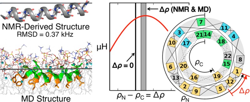
          While antimicrobial peptides (AMPs) have been widely investigated as potential therapeutics, high-resolution structures obtained under biologically relevant conditions are lacking. Here, the high-resolution structures of the homologous 22-residue long AMPs piscidin 1 (p1) and piscidin 3 (p3) are determined in fluid-phase 3:1 phosphatidylcholine/phosphatidylglycerol (PC/PG) and 1:1 phosphatidylethanolamine/phosphatidylglycerol (PE/PG) bilayers to identify molecular features important for membrane destabilization in bacterial cell membrane mimics. Structural refinement of 1H– 15N dipolar couplings and 15N chemical shifts measured by oriented sample solid-state NMR and all-atom molecular dynamics (MD) simulations provide structural and orientational information of high precision and accuracy about these interfacially bound α-helical peptides. The tilt of the helical axis, τ, is between 83° and 93° with respect to the bilayer normal for all systems and analysis methods. The average azimuthal rotation, ρ, is 235°, which results in burial of hydrophobic residues in the bilayer. The refined NMR and MD structures reveal a slight kink at G13 that delineates two helical segments characterized by a small difference in their τ angles (<10°) and significant difference in their ρ angles (∼25°). Remarkably, the kink, at the end of a G(X) 4G motif highly conserved among members of the piscidin family, allows p1 and p3 to adopt ρ angles that maximize their hydrophobic moments. Two structural features differentiate the more potent p1 from p3: p1 has a larger ρ angle and less N-terminal fraying. The peptides have comparable depths of insertion in PC/PG, but p3 is 1.2 Å more deeply inserted than p1 in PE/PG. In contrast to the ideal α-helical structures typically assumed in mechanistic models of AMPs, p1 and p3 adopt disrupted α-helical backbones that correct for differences in the amphipathicity of their N- and C-ends, and their centers of mass lie ∼1.2–3.6 Å below the plane defined by the C2 atoms of the lipid acyl chains.

          Related collections

          Most cited references72

          • Record: found
          • Abstract: found
          • Article: not found

          CHARMM-GUI: a web-based graphical user interface for CHARMM.

          CHARMM is an academic research program used widely for macromolecular mechanics and dynamics with versatile analysis and manipulation tools of atomic coordinates and dynamics trajectories. CHARMM-GUI, http://www.charmm-gui.org, has been developed to provide a web-based graphical user interface to generate various input files and molecular systems to facilitate and standardize the usage of common and advanced simulation techniques in CHARMM. The web environment provides an ideal platform to build and validate a molecular model system in an interactive fashion such that, if a problem is found through visual inspection, one can go back to the previous setup and regenerate the whole system again. In this article, we describe the currently available functional modules of CHARMM-GUI Input Generator that form a basis for the advanced simulation techniques. Future directions of the CHARMM-GUI development project are also discussed briefly together with other features in the CHARMM-GUI website, such as Archive and Movie Gallery. 2008 Wiley Periodicals, Inc.
            Bookmark
            • Record: found
            • Abstract: found
            • Article: not found

            Antimicrobial peptides of multicellular organisms.

            Multicellular organisms live, by and large, harmoniously with microbes. The cornea of the eye of an animal is almost always free of signs of infection. The insect flourishes without lymphocytes or antibodies. A plant seed germinates successfully in the midst of soil microbes. How is this accomplished? Both animals and plants possess potent, broad-spectrum antimicrobial peptides, which they use to fend off a wide range of microbes, including bacteria, fungi, viruses and protozoa. What sorts of molecules are they? How are they employed by animals in their defence? As our need for new antibiotics becomes more pressing, could we design anti-infective drugs based on the design principles these molecules teach us?
              Bookmark
              • Record: found
              • Abstract: found
              • Article: not found

              Describing the mechanism of antimicrobial peptide action with the interfacial activity model.

              Antimicrobial peptides (AMPs) have been studied for three decades, and yet a molecular understanding of their mechanism of action is still lacking. Here we summarize current knowledge for both synthetic vesicle experiments and microbe experiments, with a focus on comparisons between the two. Microbial experiments are done at peptide to lipid ratios that are at least 4 orders of magnitude higher than vesicle-based experiments. To close the gap between the two concentration regimes, we propose an "interfacial activity model", which is based on an experimentally testable molecular image of AMP-membrane interactions. The interfacial activity model may be useful in driving engineering and design of novel AMPs.
                Bookmark

                Author and article information

                Journal
                J Am Chem Soc
                J. Am. Chem. Soc
                ja
                jacsat
                Journal of the American Chemical Society
                American Chemical Society
                0002-7863
                1520-5126
                10 January 2015
                10 January 2014
                05 March 2014
                : 136
                : 9
                : 3491-3504
                Affiliations
                [] Laboratory of Computational Biology, National Heart, Lung, and Blood Institute, National Institutes of Health , Bethesda, Maryland 20892, United States
                [] Department of Chemistry and Biochemistry, University of California San Diego , La Jolla, California 92093-0307, United States
                [§ ] National High Magnetic Field Laboratory, Tallahassee, Florida 32310, United States
                [] Department of Chemistry, Hamilton College , Clinton, New York 13323, United States
                [] Department of Biological Chemistry & Molecular Pharmacology, Harvard Medical School , Boston, Massachusetts 02115, United States
                Author notes
                Article
                10.1021/ja411119m
                3985945
                24410116
                087c3f4a-c50c-458b-a632-a274cec25d37
                Copyright © 2014 American Chemical Society
                History
                : 31 October 2013
                Funding
                National Institutes of Health, United States
                Categories
                Article
                Custom metadata
                ja411119m
                ja-2013-11119m

                Chemistry
                Chemistry

                Comments

                Comment on this article