1
views
0
recommends
+1 Recommend
0 collections
    0
    shares
      • Record: found
      • Abstract: found
      • Article: found
      Is Open Access

      Meckel’s Cartilage in Mandibular Development and Dysmorphogenesis

      research-article

      Read this article at

      Bookmark
          There is no author summary for this article yet. Authors can add summaries to their articles on ScienceOpen to make them more accessible to a non-specialist audience.

          Abstract

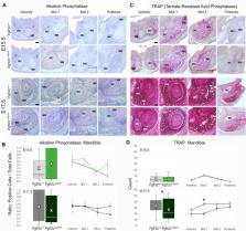
          The Fgfr2c C342Y/+ Crouzon syndrome mouse model carries a cysteine to tyrosine substitution at amino acid position 342 (Cys342Tyr; C342Y) in the fibroblast growth factor receptor 2 ( Fgfr2) gene equivalent to a FGFR2 mutation commonly associated with Crouzon and Pfeiffer syndromes in humans. The Fgfr2c C342Y mutation results in constitutive activation of the receptor and is associated with upregulation of osteogenic differentiation. Fgfr2c C342Y/+ Crouzon syndrome mice show premature closure of the coronal suture and other craniofacial anomalies including malocclusion of teeth, most likely due to abnormal craniofacial form. Malformation of the mandible can precipitate a plethora of complications including disrupting development of the upper jaw and palate, impediment of the airway, and alteration of occlusion necessary for proper mastication. The current paradigm of mandibular development assumes that Meckel’s cartilage (MC) serves as a support or model for mandibular bone formation and as a template for the later forming mandible. If valid, this implies a functional relationship between MC and the forming mandible, so mandibular dysmorphogenesis might be discerned in MC affecting the relationship between MC and mandibular bone. Here we investigate the relationship of MC to mandible development from the early mineralization of the mandible (E13.5) through the initiation of MC degradation at E17.7 using Fgfr2c C342Y/+ Crouzon syndrome embryos and their unaffected littermates ( Fgfr2c +/+ ). Differences between genotypes in both MC and mandibular bone are subtle, however MC of Fgfr2c C342Y/+ embryos is generally longer relative to unaffected littermates at E15.5 with specific aspects remaining relatively large at E17.5. In contrast, mandibular bone is smaller overall in Fgfr2c C342Y/+ embryos relative to their unaffected littermates at E15.5 with the posterior aspect remaining relatively small at E17.5. At a cellular level, differences are identified between genotypes early (E13.5) followed by reduced proliferation in MC (E15.5) and in the forming mandible (E17.5) in Fgfr2c C342Y/+ embryos. Activation of the ERK pathways is reduced in the perichondrium of MC in Fgfr2c C342Y/+ embryos and increased in bone related cells at E15.5. These data reveal that the Fgfr2c C342Y mutation differentially affects cells by type, location, and developmental age indicating a complex set of changes in the cells that make up the lower jaw.

          Related collections

          Most cited references69

          • Record: found
          • Abstract: not found
          • Book Chapter: not found

          U-Net: Convolutional Networks for Biomedical Image Segmentation

            Bookmark
            • Record: found
            • Abstract: found
            • Article: not found

            The Fibroblast Growth Factor signaling pathway

            The signaling component of the mammalian Fibroblast Growth Factor (FGF) family is comprised of eighteen secreted proteins that interact with four signaling tyrosine kinase FGF receptors (FGFRs). Interaction of FGF ligands with their signaling receptors is regulated by protein or proteoglycan cofactors and by extracellular binding proteins. Activated FGFRs phosphorylate specific tyrosine residues that mediate interaction with cytosolic adaptor proteins and the RAS-MAPK, PI3K-AKT, PLCγ, and STAT intracellular signaling pathways. Four structurally related intracellular non-signaling FGFs interact with and regulate the family of voltage gated sodium channels. Members of the FGF family function in the earliest stages of embryonic development and during organogenesis to maintain progenitor cells and mediate their growth, differentiation, survival, and patterning. FGFs also have roles in adult tissues where they mediate metabolic functions, tissue repair, and regeneration, often by reactivating developmental signaling pathways. Consistent with the presence of FGFs in almost all tissues and organs, aberrant activity of the pathway is associated with developmental defects that disrupt organogenesis, impair the response to injury, and result in metabolic disorders, and cancer. © 2015 Wiley Periodicals, Inc.
              Bookmark
              • Record: found
              • Abstract: found
              • Article: not found

              Quantification of histochemical staining by color deconvolution.

              To develop a flexible method of separation and quantification of immunohistochemical staining by means of color image analysis. An algorithm was developed to deconvolve the color information acquired with red-green-blue (RGB) cameras and to calculate the contribution of each of the applied stains based on stain-specific RGB absorption. The algorithm was tested using different combinations of diaminobenzidine, hematoxylin and eosin at different staining levels. Quantification of the different stains was not significantly influenced by the combination of multiple stains in a single sample. The color deconvolution algorithm resulted in comparable quantification independent of the stain combinations as long as the histochemical procedures did not influence the amount of stain in the sample due to bleaching because of stain solubility and saturation of staining was prevented. This image analysis algorithm provides a robust and flexible method for objective immunohistochemical analysis of samples stained with up to three different stains using a laboratory microscope, standard RGB camera setup and the public domain program NIH Image.
                Bookmark

                Author and article information

                Contributors
                Journal
                Front Genet
                Front Genet
                Front. Genet.
                Frontiers in Genetics
                Frontiers Media S.A.
                1664-8021
                16 May 2022
                2022
                : 13
                : 871927
                Affiliations
                [1] 1 Department of Anthropology , The Pennsylvania State University , University Park, PA, United States
                [2] 2 Department of Computer Science and Engineering , University of Notre Dame , Notre Dame, IN, United States
                [3] 3 Department of Genetics and Genomic Sciences , Icahn School of Medicine at Mount Sinai , New York, NY, United States
                Author notes

                Edited by: Gerson Shigeru Kobayashi, University of São Paulo, Brazil

                Reviewed by: Alice Goodwin, University of California, San Francisco, United States

                Rachel Menegaz, University of North Texas Health Science Center, United States

                *Correspondence: Joan T. Richtsmeier, jta10@ 123456psu.edu ; Susan M. Motch Perrine, qzk2@ 123456psu.edu
                [ † ]

                These authors have contributed equally to this work and share first authorship

                [ ‡ ]

                These authors have contributed equally to this work and share senior authorship

                This article was submitted to Genetics of Common and Rare Diseases, a section of the journal Frontiers in Genetics

                Article
                871927
                10.3389/fgene.2022.871927
                9149363
                0dd9ea41-fb68-435b-826b-e7d795fff460
                Copyright © 2022 Pitirri, Durham, Romano, Santos, Coupe, Zheng, Chen, Kawasaki, Jabs, Richtsmeier, Wu and Motch Perrine.

                This is an open-access article distributed under the terms of the Creative Commons Attribution License (CC BY). The use, distribution or reproduction in other forums is permitted, provided the original author(s) and the copyright owner(s) are credited and that the original publication in this journal is cited, in accordance with accepted academic practice. No use, distribution or reproduction is permitted which does not comply with these terms.

                History
                : 08 February 2022
                : 15 April 2022
                Funding
                Funded by: National Institute of Dental and Craniofacial Research , doi 10.13039/100000072;
                Award ID: R01 DE027677 R01 DE029832 R01 DE031439
                Categories
                Genetics
                Original Research

                Genetics
                craniofacial development,skull,lower jaw,mandible,embryonic cartilage,embryonic bone,crouzon syndrome,fibroblast growth factor

                Comments

                Comment on this article