36
views
0
recommends
+1 Recommend
0 collections
    0
    shares
      • Record: found
      • Abstract: found
      • Article: found
      Is Open Access

      A hydrophobic gate in the inner pore helix is the major determinant of inactivation in mechanosensitive Piezo channels

      research-article

      Read this article at

      Bookmark
          There is no author summary for this article yet. Authors can add summaries to their articles on ScienceOpen to make them more accessible to a non-specialist audience.

          Abstract

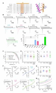
          Piezo1 and Piezo2 belong to a family of mechanically-activated ion channels implicated in a wide range of physiological processes. Mechanical stimulation triggers Piezo channels to open, but their characteristic fast inactivation process results in rapid closure. Several disease-causing mutations in Piezo1 alter the rate of inactivation, highlighting the importance of inactivation to the normal function of this channel. However, despite the structural identification of two physical constrictions within the closed pore, the mechanism of inactivation remains unknown. Here we identify a functionally conserved inactivation gate in the pore-lining inner helix of mouse Piezo1 and Piezo2 that is distinct from the two constrictions. We show that this gate controls the majority of Piezo1 inactivation via a hydrophobic mechanism and that one of the physical constrictions acts as a secondary gate. Our results suggest that, unlike other rapidly inactivating ion channels, a hydrophobic barrier gives rise to fast inactivation in Piezo channels.

          Related collections

          Most cited references49

          • Record: found
          • Abstract: found
          • Article: not found

          Piezos are pore-forming subunits of mechanically activated channels

          Mechanotransduction plays a crucial role in physiology. Biological processes including sensing touch and sound waves require yet unidentified cation channels that detect pressure. Mouse piezo1 (mpiezo1) and mpiezo2 induce mechanically activated cationic currents in cells; however, it is unknown if piezos are pore-forming ion channels or modulate ion channels. We show that Drosophila piezo (dpiezo) also induces mechanically activated currents in cells, but through channels with remarkably distinct pore properties including sensitivity to the pore blocker ruthenium red and single channel conductances. mpiezo1 assembles as a ~1.2 million-Dalton tetramer, with no evidence of other proteins in this complex. Finally, purified mpiezo1 reconstituted into asymmetric lipid bilayers and liposomes forms ruthenium red-sensitive ion channels. These data demonstrate that piezos are an evolutionarily conserved ion channel family involved in mechanotransduction.
            Bookmark
            • Record: found
            • Abstract: found
            • Article: not found

            Piezo1, a mechanically activated ion channel, is required for vascular development in mice.

            Mechanosensation is perhaps the last sensory modality not understood at the molecular level. Ion channels that sense mechanical force are postulated to play critical roles in a variety of biological processes including sensing touch/pain (somatosensation), sound (hearing), and shear stress (cardiovascular physiology); however, the identity of these ion channels has remained elusive. We previously identified Piezo1 and Piezo2 as mechanically activated cation channels that are expressed in many mechanosensitive cell types. Here, we show that Piezo1 is expressed in endothelial cells of developing blood vessels in mice. Piezo1-deficient embryos die at midgestation with defects in vascular remodeling, a process critically influenced by blood flow. We demonstrate that Piezo1 is activated by shear stress, the major type of mechanical force experienced by endothelial cells in response to blood flow. Furthermore, loss of Piezo1 in endothelial cells leads to deficits in stress fiber and cellular orientation in response to shear stress, linking Piezo1 mechanotransduction to regulation of cell morphology. These findings highlight an essential role of mammalian Piezo1 in vascular development during embryonic development.
              Bookmark
              • Record: found
              • Abstract: found
              • Article: not found

              Mechanical stretch triggers rapid epithelial cell division through Piezo1

              Despite acting as a barrier for the organs they encase, epithelial cells turnover at some of the fastest rates in the body. Yet, epithelial cell division must be tightly linked to cell death to preserve barrier function and prevent tumour formation. How do the number of dying cells match those dividing to maintain constant numbers? We previously found that when epithelial cells become too crowded, they activate the stretch-activated channel Piezo1 to trigger extrusion of cells that later die 1 . Conversely, what controls epithelial cell division to balance cell death at steady state? Here, we find that cell division occurs in regions of low cell density, where epithelial cells are stretched. By experimentally stretching epithelia, we find that mechanical stretch itself rapidly stimulates cell division through activation of the same Piezo1 channel. To do so, stretch triggers cells paused in early G2 to activate calcium-dependent ERK1/2 phosphorylation that activates cyclin B transcription necessary to drive cells into mitosis. Although both epithelial cell division and cell extrusion require Piezo1 at steady state, the type of mechanical force controls the outcome: stretch induces cell division whereas crowding induces extrusion. How Piezo1-dependent calcium transients activate two opposing processes may depend on where and how Piezo1 is activated since it accumulates in different subcellular sites with increasing cell density. In sparse epithelial regions where cells divide, Piezo1 localizes to the plasma membrane and cytoplasm whereas in dense regions where cells extrude, it forms large cytoplasmic aggregates. Because Piezo1 senses both mechanical crowding and stretch, it may act as a homeostatic sensor to control epithelial cell numbers, triggering extrusion/apoptosis in crowded regions and cell division in sparse regions.
                Bookmark

                Author and article information

                Contributors
                Role: Reviewing Editor
                Role: Senior Editor
                Journal
                eLife
                Elife
                eLife
                eLife
                eLife Sciences Publications, Ltd
                2050-084X
                10 January 2019
                2019
                : 8
                : e44003
                Affiliations
                [1 ]deptDepartment of Cellular and Molecular Physiology Yale University School of Medicine New HavenUnited States
                [2 ]deptDepartment of Neuroscience Yale University School of Medicine New HavenUnited States
                [3 ]deptProgram in Cellular Neuroscience, Neurodegeneration and Repair Yale University School of Medicine New HavenUnited States
                Universidad Nacional Autónoma de México Mexico
                The University of Texas at Austin United States
                Universidad Nacional Autónoma de México Mexico
                Author information
                http://orcid.org/0000-0001-7577-456X
                http://orcid.org/0000-0002-6661-3403
                Article
                44003
                10.7554/eLife.44003
                6349400
                30628892
                124efc6e-b0b4-4056-92b4-ba371863bdad
                © 2019, Zheng et al

                This article is distributed under the terms of the Creative Commons Attribution License, which permits unrestricted use and redistribution provided that the original author and source are credited.

                History
                : 29 November 2018
                : 10 January 2019
                Funding
                Funded by: FundRef http://dx.doi.org/10.13039/100000002, National Institutes of Health;
                Award ID: 1R01NS097547-01A1
                Award Recipient :
                Funded by: FundRef http://dx.doi.org/10.13039/100000001, National Science Foundation;
                Award ID: 1453167
                Award Recipient :
                Funded by: FundRef http://dx.doi.org/10.13039/100000002, National Institutes of Health;
                Award ID: 1R01NS091300-01A1
                Award Recipient :
                Funded by: FundRef http://dx.doi.org/10.13039/100007184, Yale School of Medicine;
                Award ID: James Hudson Brown-Alexander B Coxe Fellowship
                Award Recipient :
                The funders had no role in study design, data collection and interpretation, or the decision to submit the work for publication.
                Categories
                Research Article
                Structural Biology and Molecular Biophysics
                Custom metadata
                A functionally conserved inactivation gate in the inner helix of Piezo channels controls the majority of the inactivation process via a hydrophobic mechanism.

                Life sciences
                piezo1,piezo2,mechanosensitivity,mechanotransduction,hydrophobic gate,inactivation,none
                Life sciences
                piezo1, piezo2, mechanosensitivity, mechanotransduction, hydrophobic gate, inactivation, none

                Comments

                Comment on this article