2
views
0
recommends
+1 Recommend
0 collections
    0
    shares
      • Record: found
      • Abstract: found
      • Article: found
      Is Open Access

      TMS Correlates of Pyramidal Tract Signs and Clinical Motor Status in Patients with Cervical Spondylotic Myelopathy

      research-article

      Read this article at

      Bookmark
          There is no author summary for this article yet. Authors can add summaries to their articles on ScienceOpen to make them more accessible to a non-specialist audience.

          Abstract

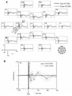
          Background: While the association between motor-evoked potential (MEP) abnormalities and motor deficit is well established, few studies have reported the correlation between MEPs and signs of pyramidal tract dysfunction without motor weakness. We assessed MEPs in patients with pyramidal signs, including motor deficits, compared to patients with pyramidal signs but without weakness. Methods: Forty-three patients with cervical spondylotic myelopathy (CSM) were dichotomized into 21 with pyramidal signs including motor deficit (Group 1) and 22 with pyramidal signs and normal strength (Group 2), and both groups were compared to 33 healthy controls (Group 0). MEPs were bilaterally recorded from the first dorsal interosseous and tibialis anterior muscle. The central motor conduction time (CMCT) was estimated as the difference between MEP latency and peripheral latency by magnetic stimulation. Peak-to-peak MEP amplitude and right-to-left differences were also measured. Results: Participants were age-, sex-, and height-matched. MEP latency in four limbs and CMCT in the lower limbs were prolonged, and MEP amplitude in the lower limbs decreased in Group 1 compared to the others. Unlike motor deficit, pyramidal signs were not associated with MEP measures, even when considering age, sex, and height as confounding factors. Conclusions: In CSM, isolated pyramidal signs may not be associated, at this stage, with MEP changes.

          Related collections

          Most cited references59

          • Record: found
          • Abstract: found
          • Article: found
          Is Open Access

          Non-invasive electrical and magnetic stimulation of the brain, spinal cord, roots and peripheral nerves: Basic principles and procedures for routine clinical and research application. An updated report from an I.F.C.N. Committee

          These guidelines provide an up-date of previous IFCN report on “Non-invasive electrical and magnetic stimulation of the brain, spinal cord and roots: basic principles and procedures for routine clinical application” (Rossini et al., 1994). A new Committee, composed of international experts, some of whom were in the panel of the 1994 “Report”, was selected to produce a current state-of-the-art review of non-invasive stimulation both for clinical application and research in neuroscience. Since 1994, the international scientific community has seen a rapid increase in non-invasive brain stimulation in studying cognition, brain–behavior relationship and pathophysiology of various neurologic and psychiatric disorders. New paradigms of stimulation and new techniques have been developed. Furthermore, a large number of studies and clinical trials have demonstrated potential therapeutic applications of non-invasive brain stimulation, especially for TMS. Recent guidelines can be found in the literature covering specific aspects of non-invasive brain stimulation, such as safety (Rossi et al., 2009), methodology (Groppa et al., 2012) and therapeutic applications (Lefaucheur et al., 2014). This up-dated review covers theoretical, physiological and practical aspects of non-invasive stimulation of brain, spinal cord, nerve roots and peripheral nerves in the light of more updated knowledge, and include some recent extensions and developments.
            Bookmark
            • Record: found
            • Abstract: not found
            • Article: not found

            NON-INVASIVE MAGNETIC STIMULATION OF HUMAN MOTOR CORTEX

              Bookmark
              • Record: found
              • Abstract: found
              • Article: not found

              TMS and drugs revisited 2014.

              The combination of pharmacology and transcranial magnetic stimulation to study the effects of drugs on TMS-evoked EMG responses (pharmaco-TMS-EMG) has considerably improved our understanding of the effects of TMS on the human brain. Ten years have elapsed since an influential review on this topic has been published in this journal (Ziemann, 2004). Since then, several major developments have taken place: TMS has been combined with EEG to measure TMS evoked responses directly from brain activity rather than by motor evoked potentials in a muscle, and pharmacological characterization of the TMS-evoked EEG potentials, although still in its infancy, has started (pharmaco-TMS-EEG). Furthermore, the knowledge from pharmaco-TMS-EMG that has been primarily obtained in healthy subjects is now applied to clinical settings, for instance, to monitor or even predict clinical drug responses in neurological or psychiatric patients. Finally, pharmaco-TMS-EMG has been applied to understand the effects of CNS active drugs on non-invasive brain stimulation induced long-term potentiation-like and long-term depression-like plasticity. This is a new field that may help to develop rationales of pharmacological treatment for enhancement of recovery and re-learning after CNS lesions. This up-dated review will highlight important knowledge and recent advances in the contribution of pharmaco-TMS-EMG and pharmaco-TMS-EEG to our understanding of normal and dysfunctional excitability, connectivity and plasticity of the human brain. Copyright © 2014 International Federation of Clinical Neurophysiology. Published by Elsevier Ireland Ltd. All rights reserved.
                Bookmark

                Author and article information

                Journal
                Brain Sci
                Brain Sci
                brainsci
                Brain Sciences
                MDPI
                2076-3425
                31 October 2020
                November 2020
                : 10
                : 11
                : 806
                Affiliations
                [1 ]Department of Surgery and Medical-Surgical Specialties, University of Catania, Via Santa Sofia, 78-95123 Catania, Italy; pennigi@ 123456unict.it
                [2 ]Department of Neurology IC, Oasi Research Institute–IRCCS, Via Conte Ruggero, 73-94018 Troina, Italy
                [3 ]Department of Neurology and Stroke Unit, ASST Cremona, Viale Concordia, 1-26100 Cremona, Italy; valentina.puglisi@ 123456asst-cremona.it (V.P.); luisa.vinciguerra@ 123456asst-cremona.it (L.V.)
                [4 ]Department of Biomedical and Biotechnological Sciences, University of Catania, Via Santa Sofia, 89-95123 Catania, Italy; drfrancescofisicaro@ 123456gmail.com (F.F.); manuela.pennisi@ 123456unict.it (M.P.)
                [5 ]Department of Neurology, San Giovanni di Dio Hospital, ASP Agrigento, Contrada Consolida, 92100 Agrigento, Italy; carlavagli@ 123456gmail.com
                [6 ]Department of Neurology, Sant’Elia Hospital, ASP Caltanissetta, Via Luigi Russo, 6-93100 Caltanissetta, Italy; m.cantone@ 123456asp.cl.it
                [7 ]Department of Medical and Surgical Sciences and Advanced Technologies, University of Catania, Via Santa Sofia, 78-95123 Catania, Italy; rbella@ 123456unict.it
                Author notes
                [* ]Correspondence: glanza@ 123456oasi.en.it ; Tel.: +39-095-3782448
                [†]

                These authors equally contributed to this work (in either order).

                [‡]

                These authors share the senior authorship (in either order).

                Author information
                https://orcid.org/0000-0002-5659-662X
                https://orcid.org/0000-0002-1579-8807
                https://orcid.org/0000-0002-9072-4971
                https://orcid.org/0000-0001-8332-6863
                Article
                brainsci-10-00806
                10.3390/brainsci10110806
                7692772
                33142762
                178367b8-0f89-4a28-96bd-6148098087c9
                © 2020 by the authors.

                Licensee MDPI, Basel, Switzerland. This article is an open access article distributed under the terms and conditions of the Creative Commons Attribution (CC BY) license ( http://creativecommons.org/licenses/by/4.0/).

                History
                : 07 September 2020
                : 26 October 2020
                Categories
                Article

                motor-evoked potentials,transcranial magnetic stimulation,corticospinal conduction,pyramidal signs,motor status,degenerative cervical myelopathy,cervical spondylotic myelopathy,clinical neuroscience

                Comments

                Comment on this article