Inviting an author to review:
Find an author and click ‘Invite to review selected article’ near their name.
Search for authorsSearch for similar articles
5
views
0
recommends
+1 Recommend
0 collections
    0
    shares
      • Record: found
      • Abstract: found
      • Article: found
      Is Open Access

      Freely Behaving Mice Can Brake and Turn During Optogenetic Stimulation of the Mesencephalic Locomotor Region

      research-article

      Read this article at

      Bookmark
          There is no author summary for this article yet. Authors can add summaries to their articles on ScienceOpen to make them more accessible to a non-specialist audience.

          Abstract

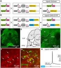
          A key function of the mesencephalic locomotor region (MLR) is to control the speed of forward symmetrical locomotor movements. However, the ability of freely moving mammals to integrate environmental cues to brake and turn during MLR stimulation is poorly documented. Here, we investigated whether freely behaving mice could brake or turn, based on environmental cues during MLR stimulation. We photostimulated the cuneiform nucleus (part of the MLR) in mice expressing channelrhodopsin in Vglut2-positive neurons in a Cre-dependent manner (Vglut2-ChR2-EYFP) using optogenetics. We detected locomotor movements using deep learning. We used patch-clamp recordings to validate the functional expression of channelrhodopsin and neuroanatomy to visualize the stimulation sites. In the linear corridor, gait diagram and limb kinematics were similar during spontaneous and optogenetic-evoked locomotion. In the open-field arena, optogenetic stimulation of the MLR evoked locomotion, and increasing laser power increased locomotor speed. Mice could brake and make sharp turns (~90°) when approaching a corner during MLR stimulation in the open-field arena. The speed during the turn was scaled with the speed before the turn, and with the turn angle. Patch-clamp recordings in Vglut2-ChR2-EYFP mice show that blue light evoked short-latency spiking in MLR neurons. Our results strengthen the idea that different brainstem neurons convey braking/turning and MLR speed commands in mammals. Our study also shows that Vglut2-positive neurons of the cuneiform nucleus are a relevant target to increase locomotor activity without impeding the ability to brake and turn when approaching obstacles, thus ensuring smooth and adaptable navigation. Our observations may have clinical relevance since cuneiform nucleus stimulation is increasingly considered to improve locomotion function in pathological states such as Parkinson’s disease, spinal cord injury, or stroke.

          Related collections

          Most cited references93

          • Record: found
          • Abstract: not found
          • Conference Proceedings: not found

          Deep Residual Learning for Image Recognition

            Bookmark
            • Record: found
            • Abstract: found
            • Article: not found

            DeepLabCut: markerless pose estimation of user-defined body parts with deep learning

            Quantifying behavior is crucial for many applications in neuroscience. Videography provides easy methods for the observation and recording of animal behavior in diverse settings, yet extracting particular aspects of a behavior for further analysis can be highly time consuming. In motor control studies, humans or other animals are often marked with reflective markers to assist with computer-based tracking, but markers are intrusive, and the number and location of the markers must be determined a priori. Here we present an efficient method for markerless pose estimation based on transfer learning with deep neural networks that achieves excellent results with minimal training data. We demonstrate the versatility of this framework by tracking various body parts in multiple species across a broad collection of behaviors. Remarkably, even when only a small number of frames are labeled (~200), the algorithm achieves excellent tracking performance on test frames that is comparable to human accuracy.
              Bookmark
              • Record: found
              • Abstract: found
              • Article: not found

              NeuN, a neuronal specific nuclear protein in vertebrates.

              A battery of monoclonal antibodies (mAbs) against brain cell nuclei has been generated by repeated immunizations. One of these, mAb A60, recognizes a vertebrate nervous system- and neuron-specific nuclear protein that we have named NeuN (Neuronal Nuclei). The expression of NeuN is observed in most neuronal cell types throughout the nervous system of adult mice. However, some major cell types appear devoid of immunoreactivity including cerebellar Purkinje cells, olfactory bulb mitral cells, and retinal photoreceptor cells. NeuN can also be detected in neurons in primary cerebellar cultures and in retinoic acid-stimulated P19 embryonal carcinoma cells. Immunohistochemically detectable NeuN protein first appears at developmental timepoints which correspond with the withdrawal of the neuron from the cell cycle and/or with the initiation of terminal differentiation of the neuron. NeuN is a soluble nuclear protein, appears as 3 bands (46-48 x 10(3) M(r)) on immunoblots, and binds to DNA in vitro. The mAb crossreacts immunohistochemically with nervous tissue from rats, chicks, humans, and salamanders. This mAb and the protein recognized by it serve as an excellent marker for neurons in the central and peripheral nervous systems in both the embryo and adult, and the protein may be important in the determination of neuronal phenotype.
                Bookmark

                Author and article information

                Contributors
                Journal
                Front Neural Circuits
                Front Neural Circuits
                Front. Neural Circuit
                Frontiers in Neural Circuits
                Frontiers Media S.A.
                1662-5110
                09 April 2021
                2021
                : 15
                : 639900
                Affiliations
                [1] 1Département de pharmacologie-physiologie, Faculté de médecine et des sciences de la santé, Université de Sherbrooke , Sherbrooke, QC, Canada
                [2] 2Department of Medical Neuroscience, Atlantic Mobility Action Project, Brain Repair Center, Dalhousie University , Halifax, NS, Canada
                [3] 3Department of Training and Movement Sciences, Humboldt-Universität zu Berlin , Berlin, Germany
                [4] 4Berlin School of Movement Science, Humboldt-Universität zu Berlin , Berlin, Germany
                [5] 5Centre de recherche du Centre hospitalier universitaire de Sherbrooke , Sherbrooke, QC, Canada
                [6] 6Centre d’excellence en neurosciences de l’Université de Sherbrooke , Sherbrooke, QC, Canada
                [7] 7Institut de pharmacologie de Sherbrooke , Sherbrooke, QC, Canada
                Author notes

                Edited by: Frederic Bretzner, Laval University, Canada

                Reviewed by: Brian R. Noga, University of Miami, United States; Patrick John Whelan, University of Calgary, Canada

                *Correspondence: Dimitri Ryczko dimitri.ryczko@ 123456usherbrooke.ca
                Article
                10.3389/fncir.2021.639900
                8062873
                33897379
                17ae19cc-7fab-45ed-ad01-e395d703849e
                Copyright © 2021 van der Zouwen, Boutin, Fougère, Flaive, Vivancos, Santuz, Akay, Sarret and Ryczko.

                This is an open-access article distributed under the terms of the Creative Commons Attribution License (CC BY). The use, distribution or reproduction in other forums is permitted, provided the original author(s) and the copyright owner(s) are credited and that the original publication in this journal is cited, in accordance with accepted academic practice. No use, distribution or reproduction is permitted which does not comply with these terms.

                History
                : 10 December 2020
                : 08 March 2021
                Page count
                Figures: 7, Tables: 0, Equations: 0, References: 94, Pages: 18, Words: 13692
                Funding
                Funded by: Canadian Institutes of Health Research 10.13039/501100000024
                Award ID: 407083 to DR, FDN-148413 to PS
                Funded by: Fonds de Recherche du Québec - Santé 10.13039/501100000156
                Award ID: 34920 to DR, 36772 to DR
                Funded by: Natural Sciences and Engineering Research Council of Canada 10.13039/501100000038
                Award ID: RTI-2019-00628 to DR, RGPIN-2017-05522 to DR
                Funded by: Canada Foundation for Innovation 10.13039/501100000196
                Categories
                Neuroscience
                Original Research

                Neurosciences
                locomotion,speed,braking,turning,mesencephalic locomotor region,cuneiform nucleus,vglut2,optogenetics

                Comments

                Comment on this article