0
views
0
recommends
+1 Recommend
0 collections
    0
    shares
      • Record: found
      • Abstract: found
      • Article: found
      Is Open Access

      Machine learning based on optimal VOI of multi-sequence MR images to predict lymphovascular invasion in invasive breast cancer

      research-article

      Read this article at

      Bookmark
          There is no author summary for this article yet. Authors can add summaries to their articles on ScienceOpen to make them more accessible to a non-specialist audience.

          Abstract

          Objectives

          Lymphovascular invasion serves as a crucial prognostic indicator in invasive breast cancer, influencing treatment decisions. We aimed to develop a machine learning model utilizing optimal volumes of interest extracted from multisequence magnetic resonance images to predict lymphovascular invasion in patients with invasive breast cancer.

          Materials and methods

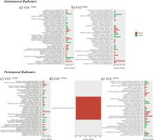
          This study comprised 191 patients postoperatively diagnosed with invasive breast cancer through multi-sequence magnetic resonance imaging. Independent predictors were identified through univariate and multivariate logistic regression analyses, culminating in the construction of a clinical model. Radiomic features were extracted from multi-sequence magnetic resonance imaging images across various volume of interest scales (−2 mm, entire, +2 mm, +4 mm, and +6 mm). Subsequently, various radiomic models were developed using machine learning model algorithms, including logistic regression, support vector machine, k-nearest neighbor, gradient boosting machine, classification and regression tree, and random forest. A hybrid model was then formulated, amalgamating optimal radiomic and clinical models.

          Results

          The area under the curve of the clinical model was 0.757. Among the radiomic models, the most efficient diagnosis was achieved by the k-nearest neighbor-based radiomics-volume of interest (+2 mm), resulting in an area under the curve of 0.780. The hybrid model, integrating the k-nearest neighbor-based radiomics-volume of interest (+2 mm), and the clinical model surpassed the individual clinical and radiomics models, exhibiting a superior area under the curve of 0.864.

          Conclusion

          Utilizing a hybrid approach integrating clinical data and multi-sequence magnetic resonance imaging-derived radiomics models based on the multiscale tumor region volume of interest (+2 mm) proved effective in determining lymphovascular invasion status in patients with invasive breast cancer. This innovative methodology may offer valuable insights for treatment planning and disease management.

          Related collections

          Most cited references35

          • Record: found
          • Abstract: found
          • Article: not found

          Global cancer statistics 2020: GLOBOCAN estimates of incidence and mortality worldwide for 36 cancers in 185 countries

          This article provides an update on the global cancer burden using the GLOBOCAN 2020 estimates of cancer incidence and mortality produced by the International Agency for Research on Cancer. Worldwide, an estimated 19.3 million new cancer cases (18.1 million excluding nonmelanoma skin cancer) and almost 10.0 million cancer deaths (9.9 million excluding nonmelanoma skin cancer) occurred in 2020. Female breast cancer has surpassed lung cancer as the most commonly diagnosed cancer, with an estimated 2.3 million new cases (11.7%), followed by lung (11.4%), colorectal (10.0 %), prostate (7.3%), and stomach (5.6%) cancers. Lung cancer remained the leading cause of cancer death, with an estimated 1.8 million deaths (18%), followed by colorectal (9.4%), liver (8.3%), stomach (7.7%), and female breast (6.9%) cancers. Overall incidence was from 2-fold to 3-fold higher in transitioned versus transitioning countries for both sexes, whereas mortality varied <2-fold for men and little for women. Death rates for female breast and cervical cancers, however, were considerably higher in transitioning versus transitioned countries (15.0 vs 12.8 per 100,000 and 12.4 vs 5.2 per 100,000, respectively). The global cancer burden is expected to be 28.4 million cases in 2040, a 47% rise from 2020, with a larger increase in transitioning (64% to 95%) versus transitioned (32% to 56%) countries due to demographic changes, although this may be further exacerbated by increasing risk factors associated with globalization and a growing economy. Efforts to build a sustainable infrastructure for the dissemination of cancer prevention measures and provision of cancer care in transitioning countries is critical for global cancer control.
            Bookmark
            • Record: found
            • Abstract: found
            • Article: not found

            Computational Radiomics System to Decode the Radiographic Phenotype

            Radiomics aims to quantify phenotypic characteristics on medical imaging through the use of automated algorithms. Radiomic artificial intelligence (AI) technology, either based on engineered hard-coded algorithms or deep learning methods, can be used to develop non-invasive imaging-based biomarkers. However, lack of standardized algorithm definitions and image processing severely hampers reproducibility and comparability of results. To address this issue, we developed PyRadiomics , a flexible open-source platform capable of extracting a large panel of engineered features from medical images. PyRadiomics is implemented in Python and can be used standalone or using 3D-Slicer. Here, we discuss the workflow and architecture of PyRadiomics and demonstrate its application in characterizing lung-lesions. Source code, documentation, and examples are publicly available at www.radiomics.io . With this platform, we aim to establish a reference standard for radiomic analyses, provide a tested and maintained resource, and to grow the community of radiomic developers addressing critical needs in cancer research.
              Bookmark
              • Record: found
              • Abstract: found
              • Article: not found

              3D Slicer as an image computing platform for the Quantitative Imaging Network.

              Quantitative analysis has tremendous but mostly unrealized potential in healthcare to support objective and accurate interpretation of the clinical imaging. In 2008, the National Cancer Institute began building the Quantitative Imaging Network (QIN) initiative with the goal of advancing quantitative imaging in the context of personalized therapy and evaluation of treatment response. Computerized analysis is an important component contributing to reproducibility and efficiency of the quantitative imaging techniques. The success of quantitative imaging is contingent on robust analysis methods and software tools to bring these methods from bench to bedside. 3D Slicer is a free open-source software application for medical image computing. As a clinical research tool, 3D Slicer is similar to a radiology workstation that supports versatile visualizations but also provides advanced functionality such as automated segmentation and registration for a variety of application domains. Unlike a typical radiology workstation, 3D Slicer is free and is not tied to specific hardware. As a programming platform, 3D Slicer facilitates translation and evaluation of the new quantitative methods by allowing the biomedical researcher to focus on the implementation of the algorithm and providing abstractions for the common tasks of data communication, visualization and user interface development. Compared to other tools that provide aspects of this functionality, 3D Slicer is fully open source and can be readily extended and redistributed. In addition, 3D Slicer is designed to facilitate the development of new functionality in the form of 3D Slicer extensions. In this paper, we present an overview of 3D Slicer as a platform for prototyping, development and evaluation of image analysis tools for clinical research applications. To illustrate the utility of the platform in the scope of QIN, we discuss several use cases of 3D Slicer by the existing QIN teams, and we elaborate on the future directions that can further facilitate development and validation of imaging biomarkers using 3D Slicer. Copyright © 2012 Elsevier Inc. All rights reserved.
                Bookmark

                Author and article information

                Contributors
                Journal
                Heliyon
                Heliyon
                Heliyon
                Elsevier
                2405-8440
                06 April 2024
                15 April 2024
                06 April 2024
                : 10
                : 7
                : e29267
                Affiliations
                [a ]Department of Radiology, The Second Affiliated Hospital of Hunan University of Chinese Medicine, Changsha, Hunan, 410005, China
                [b ]Department of Radiology, Xiangtan Central Hospital, Xiangtan, Hunan, 411100, China
                Author notes
                [* ]Corresponding author. liuhaibo521520@ 123456sina.cn
                [** ]Corresponding author. zy6645295@ 123456163.com
                [1]

                These authors contributed equally to this work.

                Article
                S2405-8440(24)05298-8 e29267
                10.1016/j.heliyon.2024.e29267
                11016709
                38623213
                1cd57b0e-9f0d-4a78-ad90-f48004a2d220
                © 2024 The Authors

                This is an open access article under the CC BY-NC license (http://creativecommons.org/licenses/by-nc/4.0/).

                History
                : 8 December 2023
                : 24 March 2024
                : 3 April 2024
                Categories
                Research Article

                invasive breast cancer,lymphovascular invasion,multi-sequence mri,machine learning,volumes of interest

                Comments

                Comment on this article