7
views
0
recommends
+1 Recommend
0 collections
    0
    shares
      • Record: found
      • Abstract: found
      • Article: found
      Is Open Access

      Contributions of the Hippocampal CA3 Circuitry to Acute Seizures and Hyperexcitability Responses in Mouse Models of Brain Ischemia

      research-article

      Read this article at

      Bookmark
          There is no author summary for this article yet. Authors can add summaries to their articles on ScienceOpen to make them more accessible to a non-specialist audience.

          Abstract

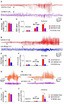
          The hippocampal circuitry is widely recognized as susceptible to ischemic injury and seizure generation. However, hippocampal contribution to acute non-convulsive seizures (NCS) in models involving middle cerebral artery occlusion (MCAO) remains to be determined. To address this, we occluded the middle cerebral artery in adult C57 black mice and monitored electroencephalographic (EEG) discharges from hippocampal and neocortical areas. Electrographic discharges in the absence of convulsive motor behaviors were observed within 90 min following occlusion of the middle cerebral artery. Hippocampal discharges were more robust than corresponding cortical discharges in all seizure events examined, and hippocampal discharges alone or with minimal cortical involvement were also observed in some seizure events. Seizure development was associated with ipsilateral hippocampal injuries as determined by subsequent histological examinations. We also introduced hypoxia-hypoglycemia episodes in mouse brain slices and examined regional hyperexcitable responses ex vivo. Extracellular recordings showed that the hippocampal CA3 region had a greater propensity for exhibiting single/multiunit activities or epileptiform field potentials following hypoxic-hypoglycemic (HH) episodes compared to the CA1, dentate gyrus, entorhinal cortical (EC) or neocortical regions. Whole-cell recordings revealed that CA3 pyramidal neurons exhibited excessive excitatory postsynaptic currents, attenuated inhibitory postsynaptic currents and intermittent or repetitive spikes in response to HH challenge. Together, these observations suggest that hippocampal discharges, possibly as a result of CA3 circuitry hyperexcitability, are a major component of acute NCS in a mouse model of MCAO.

          Related collections

          Most cited references69

          • Record: found
          • Abstract: found
          • Article: not found

          Mechanisms of spreading depression and hypoxic spreading depression-like depolarization.

          G Somjen (2001)
          Spreading depression (SD) and the related hypoxic SD-like depolarization (HSD) are characterized by rapid and nearly complete depolarization of a sizable population of brain cells with massive redistribution of ions between intracellular and extracellular compartments, that evolves as a regenerative, "all-or-none" type process, and propagates slowly as a wave in brain tissue. This article reviews the characteristics of SD and HSD and the main hypotheses that have been proposed to explain them. Both SD and HSD are composites of concurrent processes. Antagonists of N-methyl-D-aspartate (NMDA) channels or voltage-gated Na(+) or certain types of Ca(2+) channels can postpone or mitigate SD or HSD, but it takes a combination of drugs blocking all known major inward currents to effectively prevent HSD. Recent computer simulation confirmed that SD can be produced by positive feedback achieved by increase of extracellular K(+) concentration that activates persistent inward currents which then activate K(+) channels and release more K(+). Any slowly inactivating voltage and/or K(+)-dependent inward current could generate SD-like depolarization, but ordinarily, it is brought about by the cooperative action of the persistent Na(+) current I(Na,P) plus NMDA receptor-controlled current. SD is ignited when the sum of persistent inward currents exceeds persistent outward currents so that total membrane current turns inward. The degree of depolarization is not determined by the number of channels available, but by the feedback that governs the SD process. Short bouts of SD and HSD are well tolerated, but prolonged depolarization results in lasting loss of neuron function. Irreversible damage can, however, be avoided if Ca(2+) influx into neurons is prevented.
            Bookmark
            • Record: found
            • Abstract: found
            • Article: found
            Is Open Access

            Ischemic stroke: experimental models and reality

            The vast majority of cerebral stroke cases are caused by transient or permanent occlusion of a cerebral blood vessel (“ischemic stroke”) eventually leading to brain infarction. The final infarct size and the neurological outcome depend on a multitude of factors such as the duration and severity of ischemia, the existence of collateral systems and an adequate systemic blood pressure, etiology and localization of the infarct, but also on age, sex, comorbidities with the respective multimedication and genetic background. Thus, ischemic stroke is a highly complex and heterogeneous disorder. It is immediately obvious that experimental models of stroke can cover only individual specific aspects of this multifaceted disease. A basic understanding of the principal molecular pathways induced by ischemia-like conditions comes already from in vitro studies. One of the most frequently used in vivo models in stroke research is the endovascular suture or filament model in rodents with occlusion of the middle cerebral artery (MCA), which causes reproducible infarcts in the MCA territory. It does not require craniectomy and allows reperfusion by withdrawal of the occluding filament. Although promptly restored blood flow is far from the pathophysiology of spontaneous human stroke, it more closely mimics the therapeutic situation of mechanical thrombectomy which is expected to be increasingly applied to stroke patients. Direct transient or permanent occlusion of cerebral arteries represents an alternative approach but requires craniectomy. Application of endothelin-1, a potent vasoconstrictor, allows induction of transient focal ischemia in nearly any brain region and is frequently used to model lacunar stroke. Circumscribed and highly reproducible cortical lesions are characteristic of photothrombotic stroke where infarcts are induced by photoactivation of a systemically given dye through the intact skull. The major shortcoming of this model is near complete lack of a penumbra. The two models mimicking human stroke most closely are various embolic stroke models and spontaneous stroke models. Closeness to reality has its price and goes along with higher variability of infarct size and location as well as unpredictable stroke onset in spontaneous models versus unpredictable reperfusion in embolic clot models.
              Bookmark
              • Record: found
              • Abstract: found
              • Article: not found

              Closed-loop optogenetic control of thalamus as a new tool to interrupt seizures after cortical injury

              Cerebrocortical injuries, such as stroke, are a major source of disability. Maladaptive consequences can result from post-injury local reorganization of cortical circuits. For example, epilepsy is a common sequela of cortical stroke, yet mechanisms responsible for seizures following cortical injuries remain unknown. In addition to local reorganization, long-range, extra-cortical connections might be critical for seizure maintenance. Here we report in rats the first evidence that the thalamus – a structure remote from but connected to the injured cortex – is required to maintain cortical seizures. Thalamocortical neurons connected to the injured epileptic cortex undergo changes in HCN channel expression and become hyperexcitable. Targeting these neurons with a closed-loop optogenetic strategy demonstrates that reducing their activity in real-time is sufficient to immediately interrupt electrographic and behavioral seizures. This approach is of therapeutic interest for intractable epilepsy, since it spares cortical function between seizures, in contrast to existing treatments such as surgical lesioning or drugs.
                Bookmark

                Author and article information

                Contributors
                Journal
                Front Cell Neurosci
                Front Cell Neurosci
                Front. Cell. Neurosci.
                Frontiers in Cellular Neuroscience
                Frontiers Media S.A.
                1662-5102
                29 August 2018
                2018
                : 12
                : 278
                Affiliations
                [1] 1Krembil Research Institute, University Health Network , Toronto, ON, Canada
                [2] 2Department of Neurosurgery, The First Hospital of Jilin University , Changchun, China
                [3] 3Department of Medicine (Neurology), University of Toronto , Toronto, ON, Canada
                [4] 4Department of Physiology, University of Toronto , Toronto, ON, Canada
                [5] 5Department of Surgery (Neurosurgery), University of Toronto , Toronto, ON, Canada
                [6] 6Department of Neurology, The First Hospital of Jilin University , Changchun, China
                Author notes

                Edited by: Antonio Gambardella, Università degli Studi Magna Graecia di Catanzaro, Italy

                Reviewed by: Marco Fuenzalida, Universidad de Valparaíso, Chile; David Ruskin, Trinity College, United States

                *Correspondence: Jiachun Feng fengjcfrank@ 123456qq.com Liang Zhang liang.zhang@ 123456uhnresearch.ca

                These authors have contributed equally to this work

                Article
                10.3389/fncel.2018.00278
                6123792
                1fc1ea16-c8fc-471c-91de-c0c33b2761df
                Copyright © 2018 Song, Mylvaganam, Wang, Mylvaganam, Wu, Carlen, Eubanks, Feng and Zhang.

                This is an open-access article distributed under the terms of the Creative Commons Attribution License (CC BY). The use, distribution or reproduction in other forums is permitted, provided the original author(s) and the copyright owner(s) are credited and that the original publication in this journal is cited, in accordance with accepted academic practice. No use, distribution or reproduction is permitted which does not comply with these terms.

                History
                : 16 May 2018
                : 08 August 2018
                Page count
                Figures: 9, Tables: 2, Equations: 0, References: 79, Pages: 21, Words: 13508
                Categories
                Neuroscience
                Original Research

                Neurosciences
                brain slices,ca3,discharges,eeg,epileptiform,epsc,ipsc,ischemia
                Neurosciences
                brain slices, ca3, discharges, eeg, epileptiform, epsc, ipsc, ischemia

                Comments

                Comment on this article