18
views
0
recommends
+1 Recommend
0 collections
    0
    shares
      • Record: found
      • Abstract: found
      • Article: found
      Is Open Access

      Microenvironmental Regulation of Tumor Progression and Therapeutic Response in Brain Metastasis

      review-article

      Read this article at

      Bookmark
          There is no author summary for this article yet. Authors can add summaries to their articles on ScienceOpen to make them more accessible to a non-specialist audience.

          Abstract

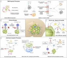
          Cellular and non-cellular components of the tumor microenvironment (TME) are emerging as key regulators of primary tumor progression, organ-specific metastasis, and therapeutic response. In the era of TME-targeted- and immunotherapies, cancer-associated inflammation has gained increasing attention. In this regard, the brain represents a unique and highly specialized organ. It has long been regarded as an immunological sanctuary site where the presence of the blood brain barrier (BBB) and blood cerebrospinal fluid barrier (BCB) restricts the entry of immune cells from the periphery. Consequently, tumor cells that metastasize to the brain were thought to be shielded from systemic immune surveillance and destruction. However, the detailed characterization of the immune landscape within border-associated areas of the central nervous system (CNS), such as the meninges and the choroid plexus, as well as the discovery of lymphatics and channels that connect the CNS with the periphery, have recently challenged the dogma of the immune privileged status of the brain. Moreover, the presence of brain metastases (BrM) disrupts the integrity of the BBB and BCB. Indeed, BrM induce the recruitment of different immune cells from the myeloid and lymphoid lineage to the CNS. Blood-borne immune cells together with brain-resident cell-types, such as astrocytes, microglia, and neurons, form a highly complex and dynamic TME that affects tumor cell survival and modulates the mode of immune responses that are elicited by brain metastatic tumor cells. In this review, we will summarize recent findings on heterotypic interactions within the brain metastatic TME and highlight specific functions of brain-resident and recruited cells at different rate-limiting steps of the metastatic cascade. Based on the insight from recent studies, we will discuss new opportunities and challenges for TME-targeted and immunotherapies for BrM.

          Related collections

          Most cited references97

          • Record: found
          • Abstract: found
          • Article: not found

          Genes that mediate breast cancer metastasis to the brain.

          The molecular basis for breast cancer metastasis to the brain is largely unknown. Brain relapse typically occurs years after the removal of a breast tumour, suggesting that disseminated cancer cells must acquire specialized functions to take over this organ. Here we show that breast cancer metastasis to the brain involves mediators of extravasation through non-fenestrated capillaries, complemented by specific enhancers of blood-brain barrier crossing and brain colonization. We isolated cells that preferentially infiltrate the brain from patients with advanced disease. Gene expression analysis of these cells and of clinical samples, coupled with functional analysis, identified the cyclooxygenase COX2 (also known as PTGS2), the epidermal growth factor receptor (EGFR) ligand HBEGF, and the alpha2,6-sialyltransferase ST6GALNAC5 as mediators of cancer cell passage through the blood-brain barrier. EGFR ligands and COX2 were previously linked to breast cancer infiltration of the lungs, but not the bones or liver, suggesting a sharing of these mediators in cerebral and pulmonary metastases. In contrast, ST6GALNAC5 specifically mediates brain metastasis. Normally restricted to the brain, the expression of ST6GALNAC5 in breast cancer cells enhances their adhesion to brain endothelial cells and their passage through the blood-brain barrier. This co-option of a brain sialyltransferase highlights the role of cell-surface glycosylation in organ-specific metastatic interactions.
            Bookmark
            • Record: found
            • Abstract: found
            • Article: not found

            Uniquely hominid features of adult human astrocytes.

            Defining the microanatomic differences between the human brain and that of other mammals is key to understanding its unique computational power. Although much effort has been devoted to comparative studies of neurons, astrocytes have received far less attention. We report here that protoplasmic astrocytes in human neocortex are 2.6-fold larger in diameter and extend 10-fold more GFAP (glial fibrillary acidic protein)-positive primary processes than their rodent counterparts. In cortical slices prepared from acutely resected surgical tissue, protoplasmic astrocytes propagate Ca(2+) waves with a speed of 36 microm/s, approximately fourfold faster than rodent. Human astrocytes also transiently increase cystosolic Ca(2+) in response to glutamatergic and purinergic receptor agonists. The human neocortex also harbors several anatomically defined subclasses of astrocytes not represented in rodents. These include a population of astrocytes that reside in layers 5-6 and extend long fibers characterized by regularly spaced varicosities. Another specialized type of astrocyte, the interlaminar astrocyte, abundantly populates the superficial cortical layers and extends long processes without varicosities to cortical layers 3 and 4. Human fibrous astrocytes resemble their rodent counterpart but are larger in diameter. Thus, human cortical astrocytes are both larger, and structurally both more complex and more diverse, than those of rodents. On this basis, we posit that this astrocytic complexity has permitted the increased functional competence of the adult human brain.
              Bookmark
              • Record: found
              • Abstract: found
              • Article: not found

              Diversity of astrocyte functions and phenotypes in neural circuits.

              Astrocytes tile the entire CNS. They are vital for neural circuit function, but have traditionally been viewed as simple, homogenous cells that serve the same essential supportive roles everywhere. Here, we summarize breakthroughs that instead indicate that astrocytes represent a population of complex and functionally diverse cells. Physiological diversity of astrocytes is apparent between different brain circuits and microcircuits, and individual astrocytes display diverse signaling in subcellular compartments. With respect to injury and disease, astrocytes undergo diverse phenotypic changes that may be protective or causative with regard to pathology in a context-dependent manner. These new insights herald the concept that astrocytes represent a diverse population of genetically tractable cells that mediate neural circuit-specific roles in health and disease.
                Bookmark

                Author and article information

                Contributors
                Journal
                Front Immunol
                Front Immunol
                Front. Immunol.
                Frontiers in Immunology
                Frontiers Media S.A.
                1664-3224
                24 July 2019
                2019
                : 10
                : 1713
                Affiliations
                [1] 1Institute for Tumor Biology and Experimental Therapy, Georg-Speyer-Haus , Frankfurt, Germany
                [2] 2Biological Sciences, Faculty 15, Goethe University , Frankfurt, Germany
                [3] 3Frankfurt Cancer Institute, Goethe University , Frankfurt, Germany
                [4] 4German Cancer Consortium (DKTK, Partner Site Frankfurt/Mainz) and German Cancer Research Center (DKFZ) , Heidelberg, Germany
                Author notes

                Edited by: Leila Akkari, Netherlands Cancer Institute (NKI), Netherlands

                Reviewed by: William K. Decker, Baylor College of Medicine, United States; Egídio Torrado, University of Minho, Portugal

                *Correspondence: Lisa Sevenich sevenich@ 123456gsh.uni-frankfurt.de

                This article was submitted to Cancer Immunity and Immunotherapy, a section of the journal Frontiers in Immunology

                †These authors have contributed equally to this work

                Article
                10.3389/fimmu.2019.01713
                6667643
                31396225
                2007d110-4b03-4d39-80ad-d328af8cb758
                Copyright © 2019 Schulz, Salamero-Boix, Niesel, Alekseeva and Sevenich.

                This is an open-access article distributed under the terms of the Creative Commons Attribution License (CC BY). The use, distribution or reproduction in other forums is permitted, provided the original author(s) and the copyright owner(s) are credited and that the original publication in this journal is cited, in accordance with accepted academic practice. No use, distribution or reproduction is permitted which does not comply with these terms.

                History
                : 15 May 2019
                : 09 July 2019
                Page count
                Figures: 2, Tables: 0, Equations: 0, References: 162, Pages: 15, Words: 13140
                Funding
                Funded by: Deutsche Krebshilfe 10.13039/501100005972
                Categories
                Immunology
                Review

                Immunology
                brain metastases,tumor microenviroment,microglia,astrocytes,immune system,immunotherapy,neurons
                Immunology
                brain metastases, tumor microenviroment, microglia, astrocytes, immune system, immunotherapy, neurons

                Comments

                Comment on this article