16
views
0
recommends
+1 Recommend
0 collections
    0
    shares
      • Record: found
      • Abstract: found
      • Article: found
      Is Open Access

      Automated detection and classification of the proximal humerus fracture by using deep learning algorithm

      research-article

      Read this article at

      Bookmark
          There is no author summary for this article yet. Authors can add summaries to their articles on ScienceOpen to make them more accessible to a non-specialist audience.

          Abstract

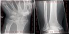
          Background and purpose — We aimed to evaluate the ability of artificial intelligence (a deep learning algorithm) to detect and classify proximal humerus fractures using plain anteroposterior shoulder radiographs.

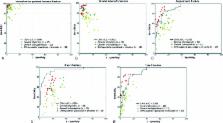
          Patients and methods — 1,891 images (1 image per person) of normal shoulders (n = 515) and 4 proximal humerus fracture types (greater tuberosity, 346; surgical neck, 514; 3-part, 269; 4-part, 247) classified by 3 specialists were evaluated. We trained a deep convolutional neural network (CNN) after augmentation of a training dataset. The ability of the CNN, as measured by top-1 accuracy, area under receiver operating characteristics curve (AUC), sensitivity/specificity, and Youden index, in comparison with humans (28 general physicians, 11 general orthopedists, and 19 orthopedists specialized in the shoulder) to detect and classify proximal humerus fractures was evaluated.

          Results — The CNN showed a high performance of 96% top-1 accuracy, 1.00 AUC, 0.99/0.97 sensitivity/specificity, and 0.97 Youden index for distinguishing normal shoulders from proximal humerus fractures. In addition, the CNN showed promising results with 65–86% top-1 accuracy, 0.90–0.98 AUC, 0.88/0.83–0.97/0.94 sensitivity/specificity, and 0.71–0.90 Youden index for classifying fracture type. When compared with the human groups, the CNN showed superior performance to that of general physicians and orthopedists, similar performance to orthopedists specialized in the shoulder, and the superior performance of the CNN was more marked in complex 3- and 4-part fractures.

          Interpretation — The use of artificial intelligence can accurately detect and classify proximal humerus fractures on plain shoulder AP radiographs. Further studies are necessary to determine the feasibility of applying artificial intelligence in the clinic and whether its use could improve care and outcomes compared with current orthopedic assessments.

          Related collections

          Most cited references7

          • Record: found
          • Abstract: not found
          • Article: not found

          Displaced proximal humeral fractures. I. Classification and evaluation.

          C S Neer (1970)
            Bookmark
            • Record: found
            • Abstract: found
            • Article: not found

            Machine learning and radiology.

            In this paper, we give a short introduction to machine learning and survey its applications in radiology. We focused on six categories of applications in radiology: medical image segmentation, registration, computer aided detection and diagnosis, brain function or activity analysis and neurological disease diagnosis from fMR images, content-based image retrieval systems for CT or MRI images, and text analysis of radiology reports using natural language processing (NLP) and natural language understanding (NLU). This survey shows that machine learning plays a key role in many radiology applications. Machine learning identifies complex patterns automatically and helps radiologists make intelligent decisions on radiology data such as conventional radiographs, CT, MRI, and PET images and radiology reports. In many applications, the performance of machine learning-based automatic detection and diagnosis systems has shown to be comparable to that of a well-trained and experienced radiologist. Technology development in machine learning and radiology will benefit from each other in the long run. Key contributions and common characteristics of machine learning techniques in radiology are discussed. We also discuss the problem of translating machine learning applications to the radiology clinical setting, including advantages and potential barriers. Copyright © 2012. Published by Elsevier B.V.
              Bookmark
              • Record: found
              • Abstract: found
              • Article: found
              Is Open Access

              Artificial intelligence for analyzing orthopedic trauma radiographs

              Background and purpose — Recent advances in artificial intelligence (deep learning) have shown remarkable performance in classifying non-medical images, and the technology is believed to be the next technological revolution. So far it has never been applied in an orthopedic setting, and in this study we sought to determine the feasibility of using deep learning for skeletal radiographs. Methods — We extracted 256,000 wrist, hand, and ankle radiographs from Danderyd’s Hospital and identified 4 classes: fracture, laterality, body part, and exam view. We then selected 5 openly available deep learning networks that were adapted for these images. The most accurate network was benchmarked against a gold standard for fractures. We furthermore compared the network’s performance with 2 senior orthopedic surgeons who reviewed images at the same resolution as the network. Results — All networks exhibited an accuracy of at least 90% when identifying laterality, body part, and exam view. The final accuracy for fractures was estimated at 83% for the best performing network. The network performed similarly to senior orthopedic surgeons when presented with images at the same resolution as the network. The 2 reviewer Cohen’s kappa under these conditions was 0.76. Interpretation — This study supports the use for orthopedic radiographs of artificial intelligence, which can perform at a human level. While current implementation lacks important features that surgeons require, e.g. risk of dislocation, classifications, measurements, and combining multiple exam views, these problems have technical solutions that are waiting to be implemented for orthopedics.
                Bookmark

                Author and article information

                Journal
                Acta Orthop
                Acta Orthop
                IORT
                iort20
                Acta Orthopaedica
                Taylor & Francis
                1745-3674
                1745-3682
                30 July 2018
                26 March 2018
                : 89
                : 4
                : 468-473
                Affiliations
                [1 ]Department of Orthopaedic Surgery and
                [2 ]Department of Dermatology , I-dermatology clinic, Seoul;
                [3 ]Department of Radiology, Konkuk University School of Medicine , Seoul;
                [4 ]Department of Orthopaedic Surgery, Kyungpook National University College of Medicine , Daegu, Korea;
                [5 ]Department of Orthopaedic Surgery, Myungji Hospital , Goyang;
                [6 ]Department of Orthopaedic Surgery, Kangwon National University College of Medicine , Chuncheon, Korea;
                [7 ]Department of Othopaedic Surgery, National Police Hospital , Seoul;
                [8 ]Department of Orthopaedic Surgery, Catholic University College of Medicine, Seoul, St Mary’s Hospital , Seoul, Korea;
                [9 ]Department of Orthopaedic Surgery, Dong-A University College of Medicine , Pusan;
                [10 ]Center for Bionics, Korea Institute of Science and Technology , Seoul, Korea
                Author notes
                Article
                1453714
                10.1080/17453674.2018.1453714
                6066766
                29577791
                226f8036-60d1-49e9-902e-e602c8db930d
                © 2018 The Author(s). Published by Taylor & Francis on behalf of the Nordic Orthopedic Federation.

                This is an Open Access article distributed under the terms of the Creative Commons Attribution-Non-Commercial License ( https://creativecommons.org/licenses/by/4.0)

                History
                : 07 January 2018
                : 21 February 2018
                Page count
                Pages: 6, Words: 4735
                Categories
                Article

                Orthopedics
                Orthopedics

                Comments

                Comment on this article