27
views
0
recommends
+1 Recommend
0 collections
    0
    shares
      • Record: found
      • Abstract: found
      • Article: found
      Is Open Access

      Age and Gender Impact the Measurement of Myocardial Interstitial Fibrosis in a Healthy Adult Chinese Population: A Cardiac Magnetic Resonance Study

      research-article

      Read this article at

      Bookmark
          There is no author summary for this article yet. Authors can add summaries to their articles on ScienceOpen to make them more accessible to a non-specialist audience.

          Abstract

          Background: Diffuse myocardial fibrosis is a common pathological process in many cardiovascular diseases. In order to determine disease, we must have standard normal imaging values. We investigated myocardial interstitial fibrosis of the left ventricle (LV) in a healthy population of Chinese adults and explored the impact of gender, age, and other physiological factors using a T1 mapping technique of cardiac magnetic resonance imaging (CMR).

          Materials and Methods: We recruited 69 healthy adult Chinese subjects (35 males; age 18–76). LV function and global strain were obtained from functional imaging. T1 mapping was performed using a modified look-locker sequence. Global and segmental native T1 and extracellular volume (ECV) were calculated using dedicated software. Gender, age, and segmental variation of both native myocardial T1 and ECV of the LV were analyzed.

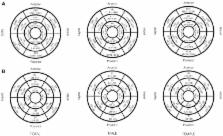
          Results: The global myocardial native T1 and ECV of the LV in this Chinese adult healthy population was 1,202 ± 45 ms and 27 ± 3% at 3T field strength, respectively. Females had a higher myocardial native T1 and ECV of the LV compared to males [1,210 (1,188–1,264) ms vs. 1,182 (1,150–1,211) ms, P < 0.001; 28 ± 3 vs. 26 ± 3%, P = 0.027, respectively]. ECV in older group was higher than younger group [27 (26–29)% vs. 25 (24–29), P = 0.019]. The multi-variate linear regression analysis showed that only gender (Beta = −0.512, P < 0.001) was independently related with global native T1 of LV while gender (Beta = −0.278, P = 0.017) and age (Beta = 0.303, P = 0.010) were independently related with global ECV of LV. From the base to apex of the LV, myocardial native T1 ( P = 0.020) and ECV ( P < 0.001) significantly increased. Within the same slice of the LV, there were significant segmental variations of both myocardial native T1 ( P < 0.001) and ECV ( P < 0.001) values.

          Conclusion: Gender and age have significant impacts on the imaging markers of myocardial interstitial fibrosis in healthy adult Chinese volunteers. Segmental variation of myocardial interstitial fibrosis was also observed.

          Related collections

          Most cited references15

          • Record: found
          • Abstract: found
          • Article: not found

          Extracellular volume imaging by magnetic resonance imaging provides insights into overt and sub-clinical myocardial pathology.

          Conventional late gadolinium enhancement (LGE) cardiac magnetic resonance can detect myocardial infarction and some forms of non-ischaemic myocardial fibrosis. However, quantitative imaging of extracellular volume fraction (ECV) may be able to detect subtle abnormalities such as diffuse fibrosis or post-infarct remodelling of remote myocardium. The aims were (1) to measure ECV in myocardial infarction and non-ischaemic myocardial fibrosis, (2) to determine whether ECV varies with age, and (3) to detect sub-clinical abnormalities in 'normal appearing' myocardium remote from regions of infarction. Cardiac magnetic resonance ECV imaging was performed in 126 patients with T1 mapping before and after injection of gadolinium contrast. Conventional LGE images were acquired for the left ventricle. In patients with a prior myocardial infarction, the infarct region had an ECV of 51 ± 8% which did not overlap with the remote 'normal appearing' myocardium that had an ECV of 27 ± 3% (P < 0.001, n = 36). In patients with non-ischaemic cardiomyopathy, the ECV of atypical LGE was 37 ± 6%, whereas the 'normal appearing' myocardium had an ECV of 26 ± 3% (P < 0.001, n = 30). The ECV of 'normal appearing' myocardium increased with age (r = 0.28, P = 0.01, n = 60). The ECV of 'normal appearing' myocardium remote from myocardial infarctions increased as left ventricular ejection fraction decreased (r = -0.50, P = 0.02). Extracellular volume fraction imaging can quantitatively characterize myocardial infarction, atypical diffuse fibrosis, and subtle myocardial abnormalities not clinically apparent on LGE images. Taken within the context of prior literature, these subtle ECV abnormalities are consistent with diffuse fibrosis related to age and changes remote from infarction.
            Bookmark
            • Record: found
            • Abstract: found
            • Article: found
            Is Open Access

            Aging and the cardiac collagen matrix: Novel mediators of fibrotic remodelling

            Cardiovascular disease is a leading cause of death worldwide and there is a pressing need for new therapeutic strategies to treat such conditions. The risk of developing cardiovascular disease increases dramatically with age, yet the majority of experimental research is executed using young animals. The cardiac extracellular matrix (ECM), consisting predominantly of fibrillar collagen, preserves myocardial integrity, provides a means of force transmission and supports myocyte geometry. Disruptions to the finely balanced control of collagen synthesis, post-synthetic deposition, post-translational modification and degradation may have detrimental effects on myocardial functionality. It is now well established that the aged heart is characterized by fibrotic remodelling, but the mechanisms responsible for this are incompletely understood. Furthermore, studies using aged animal models suggest that interstitial remodelling with disease may be age-dependent. Thus with the identification of new therapeutic strategies targeting fibrotic remodelling, it may be necessary to consider age-dependent mechanisms. In this review, we discuss remodelling of the cardiac collagen matrix as a function of age, whilst highlighting potential novel mediators of age-dependent fibrotic pathways.
              Bookmark
              • Record: found
              • Abstract: found
              • Article: not found

              Gender differences and aging: effects on the human heart.

              This study investigated the changes in myocyte size and number in the left and right ventricles that occur with aging in the female and male heart. Differences in life span between women and men may be related to a better preservation of myocardial structure in the female heart with aging. On this basis, the hypothesis was advanced that the aging process has a different impact on the integrity of the myocardium in the two genders. Morphometric methodologies were applied to analyze the changes in number and size of ventricular myocytes in the hearts of 53 women and 53 men. The changes in mononucleated and binucleated myocytes with age were determined in enzymatically dissociated cells. The age interval examined varied from 17 to 95 years. Aging was associated with a preservation of ventricular myocardial mass, aggregate number of mononucleated and binucleated myocytes, average cell diameter and volume in the female heart. In contrast, nearly 1 g/year of myocardium was lost in the male heart, and this phenomenon accounted for the loss of approximately 64 million cells. This detrimental effect involved the left and right sides of the heart. In the remaining cells, myocyte cell volume increased at a rate of 158 microns3/year in the left and 167 microns3/year in the right ventricle. Aging does not lead to myocyte cell loss and myocyte cellular reactive hypertrophy in women, indicating that gender differences may play a significant role in the detrimental effects of the aging process on the heart.
                Bookmark

                Author and article information

                Contributors
                Journal
                Front Physiol
                Front Physiol
                Front. Physiol.
                Frontiers in Physiology
                Frontiers Media S.A.
                1664-042X
                06 March 2018
                2018
                : 9
                : 140
                Affiliations
                [1] 1Cardiology Division, West China Hospital, Sichuan University , Chengdu, China
                [2] 2Cardiovascular Division, Department of Medicine, University of Pennsylvania , Philadelphia, PA, United States
                [3] 3Radiology Department, West China Hospital, Sichuan University , Chengdu, China
                [4] 4Siemens Healthcare GmbH , Erlangen, Germany
                [5] 5Northeast Asia MR Collaboration, Siemens Healthcare , Beijing, China
                Author notes

                Edited by: Jerzy Sacha, Opole University of Technology, Poland

                Reviewed by: Jarna Hannukainen, University of Turku, Finland; Ilkka H. A. Heinonen, University of Turku, Finland; Alessandro Cannavo, Temple University, United States

                *Correspondence: Yucheng Chen chenyucheng2003@ 123456126.com

                This article was submitted to Clinical and Translational Physiology, a section of the journal Frontiers in Physiology

                †These authors have contributed equally to this work.

                Article
                10.3389/fphys.2018.00140
                5845542
                29559916
                23906682-9f01-4933-97c1-fe797645498a
                Copyright © 2018 Dong, Yang, Han, Cheng, Sun, Wan, Liu, Greiser, Zhou and Chen.

                This is an open-access article distributed under the terms of the Creative Commons Attribution License (CC BY). The use, distribution or reproduction in other forums is permitted, provided the original author(s) and the copyright owner are credited and that the original publication in this journal is cited, in accordance with accepted academic practice. No use, distribution or reproduction is permitted which does not comply with these terms.

                History
                : 28 March 2017
                : 12 February 2018
                Page count
                Figures: 2, Tables: 4, Equations: 0, References: 33, Pages: 9, Words: 6336
                Funding
                Funded by: National Natural Science Foundation of China 10.13039/501100001809
                Award ID: No. 81271531
                Award ID: No. 81571638
                Categories
                Physiology
                Original Research

                Anatomy & Physiology
                myocardial interstitial fibrosis,t1 mapping,native t1,extracellular volume (ecv),gender,age,chinese population

                Comments

                Comment on this article