9
views
0
recommends
+1 Recommend
0 collections
    0
    shares
      • Record: found
      • Abstract: found
      • Article: found
      Is Open Access

      Single cell correlation analysis of liquid and solid biopsies in metastatic colorectal cancer

      research-article

      Read this article at

      Bookmark
          There is no author summary for this article yet. Authors can add summaries to their articles on ScienceOpen to make them more accessible to a non-specialist audience.

          Abstract

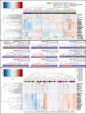
          As cancer care is transitioning to personalized therapies with necessary complementary or companion biomarkers there is significant interest in determining to what extent non-invasive liquid biopsies reflect the gold standard solid biopsy. We have established an approach for measuring patient-specific circulating and solid cell concordance by introducing tumor touch preparations to the High-Definition Single Cell Analysis workflow for high-resolution cytomorphometric characterization of metastatic colorectal cancer (mCRC). Subgroups of cells based on size, shape and protein expression were identified in both liquid and solid biopsies, which overall displayed high inter- and intra- patient pleomorphism at the single-cell level of analysis. Concordance of liquid and solid biopsies was patient-dependent and between 0.1-0.9. Morphometric variables displayed particularly high correlation, suggesting that circulating cells do not represent distinct subpopulations from the solid tumor. This was further substantiated by significant decrease in concentration of circulating cells after mCRC resection. Combined with the association of circulating cells with tumor burden and necrosis of hepatic lesions, our overall findings demonstrate that liquid biopsy cells can be informative biomarkers in the mCRC setting. Patient-specific level of concordance can readily be measured to establish the utility of circulating cells as biomarkers and define biosignatures for liquid biopsy assays.

          Related collections

          Most cited references25

          • Record: found
          • Abstract: found
          • Article: not found

          Actual 10-year survival after resection of colorectal liver metastases defines cure.

          Resection of colorectal liver metastases (CLM) in selected patients has evolved as the standard of care during the last 20 years. In the absence of prospective randomized clinical trials, a survival benefit has been deduced relative to historical controls based on actuarial data. There is now sufficient follow-up on a significant number of patients to address the curative intent of resecting CLM. Retrospective review of a prospectively maintained database was performed on patients who underwent resection of CLM from 1985 to 1994. Postoperative deaths were excluded. Disease-specific survival (DSS) was calculated from the time of hepatectomy using the Kaplan-Meier method. There were 612 consecutive patients identified with 10-year follow-up. Median DSS was 44 months. There were 102 actual 10-year survivors. Ninety-nine (97%) of the 102 were disease free at last follow-up. Only one patient experienced a disease-specific death after 10 years of survival. In contrast, 34% of the 5-year survivors suffered a cancer-related death. Previously identified poor prognostic factors found among the 102 actual 10-year survivors included 7% synchronous disease, 36% disease-free interval less than 12 months, 25% bilobar metastases, 50% node-positive primary, 39% more than one metastasis, and 35% tumor size more than 5 cm. Patients who survive 10 years appear to be cured of their disease, whereas approximately one third of actual 5-year survivors succumb to a cancer-related death. In well-selected patients, there is at least a one in six chance of cure after hepatectomy for CLM. The presence of poor prognostic factors does not preclude the possibility of long-term survival and cure.
            Bookmark
            • Record: found
            • Abstract: found
            • Article: found
            Is Open Access

            Significance of Circulating Tumor Cells Detected by the CellSearch System in Patients with Metastatic Breast Colorectal and Prostate Cancer

            The increasing number of treatment options for patients with metastatic carcinomas has created a concomitant need for new methods to monitor their use. Ideally, these modalities would be noninvasive, be independent of treatment, and provide quantitative real-time analysis of tumor activity in a variety of carcinomas. Assessment of circulating tumor cells (CTCs) shed into the blood during metastasis may satisfy this need. We developed the CellSearch System to enumerate CTC from 7.5 mL of venous blood. In this review we compare the outcomes from three prospective multicenter studies investigating the use of CTC to monitor patients undergoing treatment for metastatic breast (MBC), colorectal (MCRC), or prostate cancer (MPC) and review the CTC definition used in these studies. Evaluation of CTC at anytime during the course of disease allows assessment of patient prognosis and is predictive of overall survival.
              Bookmark
              • Record: found
              • Abstract: found
              • Article: not found

              Circulating Tumor Cells Undergoing EMT Provide a Metric for Diagnosis and Prognosis of Patients with Hepatocellular Carcinoma

              To clarify the significance of circulating tumor cells (CTC) undergoing epithelial-mesenchymal transition (EMT) in patients with hepatocellular carcinoma (HCC), we used an advanced CanPatrol CTC-enrichment technique and in situ hybridization to enrich and classify CTC from blood samples. One hundred and one of 112 (90.18%) patients with HCC were CTC positive, even with early-stage disease. CTCs were also detected in 2 of 12 patients with hepatitis B virus (HBV), both of whom had small HCC tumors detected within 5 months. CTC count ≥16 and mesenchymal-CTC (M-CTC) percentage ≥2% prior to resection were significantly associated with early recurrence, multi-intrahepatic recurrence, and lung metastasis. Postoperative CTC monitoring in 10 patients found that most had an increased CTC count and M-CTC percentage before clinically detectable recurrence nodules appeared. Analysis of HCC with high CTC count and high M-CTC percentage identified 67 differentially expressed cancer-related genes involved in cancer-related biological pathways (e.g., cell adhesion and migration, tumor angiogenesis, and apoptosis). One of the identified genes, BCAT1, was significantly upregulated, and knockdown in Hepg2, Hep3B, and Huh7 cells reduced cell proliferation, migration, and invasion while promoting apoptosis. A concomitant increase in epithelial marker expression (EpCAM and E-cadherin) and reduced mesenchymal marker expression (vimentin and Twist) suggest that BCAT1 may trigger the EMT process. Overall, CTCs were highly correlated with HCC characteristics, representing a novel marker for early diagnosis and a prognostic factor for early recurrence. BCAT1 overexpression may induce CTC release by triggering EMT and may be an important biomarker of HCC metastasis.Significance: In liver cancer, CTC examination may represent an important "liquid biopsy" tool to detect both early disease and recurrent or metastatic disease, providing cues for early intervention or adjuvant therapy. Cancer Res; 78(16); 4731-44. ©2018 AACR.
                Bookmark

                Author and article information

                Journal
                Oncotarget
                Oncotarget
                Oncotarget
                ImpactJ
                Oncotarget
                Impact Journals LLC
                1949-2553
                17 December 2019
                17 December 2019
                : 10
                : 66
                : 7016-7030
                Affiliations
                1USC Michelson Center for Convergent Bioscience, University of Southern California, Los Angeles, CA, USA
                2Biomedical Center, Faculty of Medicine in Pilsen, Charles University, Pilsen, Czech Republic
                3Scripps MDAnderson Cancer Center, La Jolla, CA, USA
                4Division of Surgical Oncology, Baylor College of Medicine, Houston, TX, USA
                5Keck School of Medicine, University of Southern California, Los Angeles, CA, USA
                6Diagnostic and Experimental Pathology, Eli Lilly and Company, Lilly Corporate Center, Indianapolis, IN, USA
                Author notes
                Correspondence to: Peter Kuhn, pkuhn@ 123456usc.edu
                Article
                27271
                10.18632/oncotarget.27271
                6925029
                239ca065-f41e-435f-94a9-dd689a560fdf
                Copyright: © 2019 Gerdtsson et al.

                This is an open-access article distributed under the terms of the Creative Commons Attribution License 3.0 (CC BY 3.0), which permits unrestricted use, distribution, and reproduction in any medium, provided the original author and source are credited.

                History
                : 10 July 2019
                : 24 September 2019
                Categories
                Research Paper

                Oncology & Radiotherapy
                single cell analysis,liquid biopsy,metastatic colorectal cancer,circulating tumor cells,cytomorphology

                Comments

                Comment on this article