144
views
0
recommends
+1 Recommend
0 collections
    12
    shares
      • Record: found
      • Abstract: found
      • Article: found
      Is Open Access

      The Tumor Suppressors p53, p63, and p73 Are Regulators of MicroRNA Processing Complex

      research-article
      *
      PLoS ONE
      Public Library of Science

      Read this article at

      Bookmark
          There is no author summary for this article yet. Authors can add summaries to their articles on ScienceOpen to make them more accessible to a non-specialist audience.

          Abstract

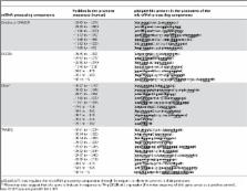
          The tumor suppressors p53, p73, and p63 are known to function as transcription factors. They promote either growth arrest or apoptosis, depending upon the DNA damage. A number of microRNAs (miRNAs) have been shown to function as transcriptional targets of p53 and they appear to aid p53 in promoting growth arrest and apoptosis. However, the question of p53/p63/p73 regulating the miRNA processing complex has not been addressed in depth so far. Comparative/computational genomic analysis was performed using Target scan, Mami, and Diana software to identify miRNAs that regulate the miRNA processing complex. Here, I present evidence for the first time that the tumor suppressors p53, p63, and p73 function as both positive and negative regulators of the miRNA processing components. Curated p53-dependent miRNA expression data was used to identify p53-miRs that target the components of the miRNA-processing complex. This analysis suggests that most of the components (mRNAs' 3′UTR) of the miRNA processing complex are targeted by p53-miRs. Remarkably, this data revealed the conserved nature of p53-miRs in targeting a number of components of the miRNA processing complex. p53/p73/p63 appears to regulate the major components of the miRNA processing, such as Drosha-DGCR8, Dicer-TRBP2, and Argonaute proteins. In particular, p53/p73/p63 appears to regulate the processing of miRNAs, such as let-7, miR-200c, miR-143, miR-107, miR-16, miR-145, miR-134, miR-449a, miR-503, and miR-21. Interestingly, there seems to be a phenotypic similarity between p63 −/− and dicer −/− mice, suggesting that p63 and dicer could regulate each other. In addition, p63, p73, and the DGCR8 proteins contain a conserved interaction domain. Further, promoters of a number of components of the miRNA processing machinery, including dicer and P2P-R, contain p53-REs, suggesting that they could be direct transcriptional targets of p63/p73/p53. Together, this study provides mechanistic insights into how p53, p63, and p73 regulate the components of the miRNA processing; and how p53, TA-p63, and TA-p73 regulated miRNAs inhibit tumorigenesis, EMT, metastasis, and cancer stem cell proliferation.

          Related collections

          Most cited references61

          • Record: found
          • Abstract: found
          • Article: not found

          Switching from repression to activation: microRNAs can up-regulate translation.

          AU-rich elements (AREs) and microRNA target sites are conserved sequences in messenger RNA (mRNA) 3' untranslated regions (3'UTRs) that control gene expression posttranscriptionally. Upon cell cycle arrest, the ARE in tumor necrosis factor-alpha (TNFalpha) mRNA is transformed into a translation activation signal, recruiting Argonaute (AGO) and fragile X mental retardation-related protein 1 (FXR1), factors associated with micro-ribonucleoproteins (microRNPs). We show that human microRNA miR369-3 directs association of these proteins with the AREs to activate translation. Furthermore, we document that two well-studied microRNAs-Let-7 and the synthetic microRNA miRcxcr4-likewise induce translation up-regulation of target mRNAs on cell cycle arrest, yet they repress translation in proliferating cells. Thus, activation is a common function of microRNPs on cell cycle arrest. We propose that translation regulation by microRNPs oscillates between repression and activation during the cell cycle.
            Bookmark
            • Record: found
            • Abstract: found
            • Article: not found

            SMAD proteins control DROSHA-mediated microRNA maturation.

            MicroRNAs (miRNAs) are small non-coding RNAs that participate in the spatiotemporal regulation of messenger RNA and protein synthesis. Aberrant miRNA expression leads to developmental abnormalities and diseases, such as cardiovascular disorders and cancer; however, the stimuli and processes regulating miRNA biogenesis are largely unknown. The transforming growth factor beta (TGF-beta) and bone morphogenetic protein (BMP) family of growth factors orchestrates fundamental biological processes in development and in the homeostasis of adult tissues, including the vasculature. Here we show that induction of a contractile phenotype in human vascular smooth muscle cells by TGF-beta and BMPs is mediated by miR-21. miR-21 downregulates PDCD4 (programmed cell death 4), which in turn acts as a negative regulator of smooth muscle contractile genes. Surprisingly, TGF-beta and BMP signalling promotes a rapid increase in expression of mature miR-21 through a post-transcriptional step, promoting the processing of primary transcripts of miR-21 (pri-miR-21) into precursor miR-21 (pre-miR-21) by the DROSHA (also known as RNASEN) complex. TGF-beta- and BMP-specific SMAD signal transducers are recruited to pri-miR-21 in a complex with the RNA helicase p68 (also known as DDX5), a component of the DROSHA microprocessor complex. The shared cofactor SMAD4 is not required for this process. Thus, regulation of miRNA biogenesis by ligand-specific SMAD proteins is critical for control of the vascular smooth muscle cell phenotype and potentially for SMAD4-independent responses mediated by the TGF-beta and BMP signalling pathways.
              Bookmark
              • Record: found
              • Abstract: found
              • Article: not found

              Relief of microRNA-mediated translational repression in human cells subjected to stress.

              In metazoans, most microRNAs imperfectly base-pair with the 3' untranslated region (3'UTR) of target mRNAs and prevent protein accumulation by either repressing translation or inducing mRNA degradation. Examples of specific mRNAs undergoing microRNA-mediated repression are numerous, but whether the repression is a reversible process remains largely unknown. Here we show that cationic amino acid transporter 1 (CAT-1) mRNA and reporters bearing its 3'UTR can be relieved from the microRNA miR-122-induced inhibition in human hepatocarcinoma cells subjected to different stress conditions. The derepression of CAT-1 mRNA is accompanied by its release from cytoplasmic processing bodies and its recruitment to polysomes. The derepression requires binding of HuR, an AU-rich-element binding protein, to the 3'UTR of CAT-1 mRNA. We propose that proteins interacting with the 3'UTR will generally act as modifiers altering the potential of miRNAs to repress gene expression.
                Bookmark

                Author and article information

                Contributors
                Role: Editor
                Journal
                PLoS One
                plos
                plosone
                PLoS ONE
                Public Library of Science (San Francisco, USA )
                1932-6203
                2010
                12 May 2010
                : 5
                : 5
                : e10615
                Affiliations
                [1]Genome Discovery, Murungapakkam, India
                Roswell Park Cancer Institute, United States of America
                Author notes

                Conceived and designed the experiments: LPB. Performed the experiments: LPB. Analyzed the data: LPB. Contributed reagents/materials/analysis tools: LPB. Wrote the paper: LPB.

                Article
                10-PONE-RA-15501
                10.1371/journal.pone.0010615
                2868896
                20485546
                246f9d26-9fff-4eda-8816-913259087756
                Lakshmanane Boominathan. This is an open-access article distributed under the terms of the Creative Commons Attribution License, which permits unrestricted use, distribution, and reproduction in any medium, provided the original author and source are credited.
                History
                : 13 January 2010
                : 18 March 2010
                Page count
                Pages: 13
                Categories
                Research Article
                Oncology
                Cell Biology/Cell Growth and Division
                Cell Biology/Cell Signaling
                Cell Biology/Cellular Death and Stress Responses
                Cell Biology/Gene Expression

                Uncategorized
                Uncategorized

                Comments

                Comment on this article