39
views
0
recommends
+1 Recommend
0 collections
    0
    shares
      • Record: found
      • Abstract: found
      • Article: found
      Is Open Access

      Macrophagic and microglial responses after focal traumatic brain injury in the female rat

      research-article

      Read this article at

      Bookmark
          There is no author summary for this article yet. Authors can add summaries to their articles on ScienceOpen to make them more accessible to a non-specialist audience.

          Abstract

          Background

          After central nervous system injury, inflammatory macrophages (M1) predominate over anti-inflammatory macrophages (M2). The temporal profile of M1/M2 phenotypes in macrophages and microglia after traumatic brain injury (TBI) in rats is unknown. We subjected female rats to severe controlled cortical impact (CCI) and examined the postinjury M1/M2 time course in their brains.

          Methods

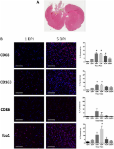
          The motor cortex (2.5 mm left laterally and 1.0 mm anteriorly from the bregma) of anesthetized female Wistar rats (ages 8 to 10 weeks; N = 72) underwent histologically moderate to severe CCI with a 5-mm impactor tip. Separate cohorts of rats had their brains dissociated into cells for flow cytometry, perfusion-fixed for immunohistochemistry (IHC) and ex vivo magnetic resonance imaging or flash-frozen for RNA and protein analysis. For each analytical method used, separate postinjury times were included for 24 hours; 3 or 5 days; or 1, 2, 4 or 8 weeks.

          Results

          By IHC, we found that the macrophagic and microglial responses peaked at 5 to 7 days post-TBI with characteristics of mixed populations of M1 and M2 phenotypes. Upon flow cytometry examination of immunological cells isolated from brain tissue, we observed that peak M2-associated staining occurred at 5 days post-TBI. Chemokine analysis by multiplex assay showed statistically significant increases in macrophage inflammatory protein 1α and keratinocyte chemoattractant/growth-related oncogene on the ipsilateral side within the first 24 hours after injury relative to controls and to the contralateral side. Quantitative RT-PCR analysis demonstrated expression of both M1- and M2-associated markers, which peaked at 5 days post-TBI.

          Conclusions

          The responses of macrophagic and microglial cells to histologically severe CCI in the female rat are maximal between days 3 and 7 postinjury. The response to injury is a mixture of M1 and M2 phenotypes.

          Related collections

          Most cited references45

          • Record: found
          • Abstract: found
          • Article: not found

          Heterogeneity of Microglial Activation in the Innate Immune Response in the Brain

          The immune response in the brain has been widely investigated and while many studies have focused on the proinflammatory cytotoxic response, the brain’s innate immune system demonstrates significant heterogeneity. Microglia, like other tissue macrophages, participate in repair and resolution processes after infection or injury to restore normal tissue homeostasis. This review examines the mechanisms that lead to reduction of self-toxicity and to repair and restructuring of the damaged extracellular matrix in the brain. Part of the resolution process involves switching macrophage functional activation to include reduction of proinflammatory mediators, increased production and release of anti-inflammatory cytokines, and production of cytoactive factors involved in repair and reconstruction of the damaged brain. Two partially overlapping and complimentary functional macrophage states have been identified and are called alternative activation and acquired deactivation. The immunosuppressive and repair processes of each of these states and how alternative activation and acquired deactivation participate in chronic neuroinflammation in the brain are discussed.
            Bookmark
            • Record: found
            • Abstract: found
            • Article: not found

            Macrophages sequentially change their functional phenotype in response to changes in microenvironmental influences.

            Recent studies have described the development of distinct functional subsets of macrophages in association with cancer, autoimmune disease, and chronic infections. Based on the ability of Th1 vs Th2 cytokines to promote opposing activities in macrophages, it has been proposed that macrophages develop into either type 1 inflammatory or type 2 anti-inflammatory subsets. As an alternative to the concept of subset development, we propose that macrophages, in response to changes in their tissue environment, can reversibly and progressively change the pattern of functions that they express. As demonstrated herein, macrophages can reversibly shift their functional phenotype through a multitude of patterns in response to changes in cytokine environment. Macrophages display distinct functional patterns after treatment with IFN-gamma, IL-12, IL-4, or IL-10 and additional functional patterns are displayed depending on whether the cytokine is present alone or with other cytokines and whether the cytokines are added before or concomitantly with the activating stimulus (LPS). Sequential treatment of macrophages with multiple cytokines results in a progression through multiple functional phenotypes. This ability to adapt to changing cytokine environments has significant in vivo relevance, as evidenced by the demonstration that macrophage functional phenotypes established in vivo in aged or tumor-bearing mice can be altered by changing their microenvironment. A concept of functional adaptivity is proposed that has important implications for therapeutic targeting of macrophages in chronic diseases that result in the dominance of particular functional phenotypes of macrophages that play a significant role in disease pathology.
              Bookmark
              • Record: found
              • Abstract: found
              • Article: not found

              Neuroinflammation after traumatic brain injury: opportunities for therapeutic intervention.

              Traumatic brain injury (TBI) remains one of the leading causes of mortality and morbidity worldwide, yet despite extensive efforts to develop neuroprotective therapies for this devastating disorder there have been no successful outcomes in human clinical trials to date. Following the primary mechanical insult TBI results in delayed secondary injury events due to neurochemical, metabolic and cellular changes that account for many of the neurological deficits observed after TBI. The development of secondary injury represents a window of opportunity for therapeutic intervention to prevent progressive tissue damage and loss of function after injury. To establish effective neuroprotective treatments for TBI it is essential to fully understand the complex cellular and molecular events that contribute to secondary injury. Neuroinflammation is well established as a key secondary injury mechanism after TBI, and it has been long considered to contribute to the damage sustained following brain injury. However, experimental and clinical research indicates that neuroinflammation after TBI can have both detrimental and beneficial effects, and these likely differ in the acute and delayed phases after injury. The key to developing future anti-inflammatory based neuroprotective treatments for TBI is to minimize the detrimental and neurotoxic effects of neuroinflammation while promoting the beneficial and neurotrophic effects, thereby creating optimal conditions for regeneration and repair after injury. This review outlines how post-traumatic neuroinflammation contributes to secondary injury after TBI, and discusses the complex and varied responses of the primary innate immune cells of the brain, microglia, to injury. In addition, emerging experimental anti-inflammatory and multipotential drug treatment strategies for TBI are discussed, as well as some of the challenges faced by the research community to translate promising neuroprotective drug treatments to the clinic. Copyright © 2012 Elsevier Inc. All rights reserved.
                Bookmark

                Author and article information

                Contributors
                Journal
                J Neuroinflammation
                J Neuroinflammation
                Journal of Neuroinflammation
                BioMed Central
                1742-2094
                2014
                24 April 2014
                : 11
                : 82
                Affiliations
                [1 ]Center for Neuroscience and Regenerative Medicine, Uniformed Services University of the Health Sciences, 4301 Jones Bridge Road, Bethesda, MD 20814, USA
                [2 ]Frank Laboratory, National Institutes of Health, Building 10, Room B1N256, MSC 1074, 10 Center Drive, Bethesda, MD 20814, USA
                [3 ]National Institute of Biomedical Imaging and Bioengineering, National Institutes of Health, 10 Center Drive, Bethesda, MD 20814, USA
                [4 ]Department of Environmental Science, Baylor University, One Bear Place #97266, Waco, TX 76798, USA
                [5 ]Department of Neurosurgery, Medical College of Wisconsin, 8701 Watertown Plank Road, Milwaukee, WI 53226, USA
                Article
                1742-2094-11-82
                10.1186/1742-2094-11-82
                4022366
                24761998
                247bef5f-0fc7-4ad2-ac77-02ef94191128
                Copyright © 2014 Turtzo et al.; licensee BioMed Central Ltd.

                This is an Open Access article distributed under the terms of the Creative Commons Attribution License ( http://creativecommons.org/licenses/by/4.0), which permits unrestricted use, distribution, and reproduction in any medium, provided the original work is properly credited. The Creative Commons Public Domain Dedication waiver ( http://creativecommons.org/publicdomain/zero/1.0/) applies to the data made available in this article, unless otherwise stated.

                History
                : 3 February 2014
                : 6 April 2014
                Categories
                Research

                Neurosciences
                controlled cortical impact,inflammation,macrophage,microglia,mri,rat,traumatic brain injury
                Neurosciences
                controlled cortical impact, inflammation, macrophage, microglia, mri, rat, traumatic brain injury

                Comments

                Comment on this article