2
views
0
recommends
+1 Recommend
0 collections
    0
    shares
      • Record: found
      • Abstract: found
      • Article: found
      Is Open Access

      Rodent Model of Muscular Atrophy for Sarcopenia Study

      review-article

      Read this article at

      Bookmark
          There is no author summary for this article yet. Authors can add summaries to their articles on ScienceOpen to make them more accessible to a non-specialist audience.

          Abstract

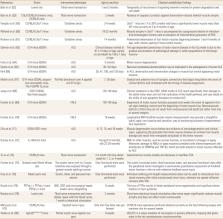
          The hallmark symptom of sarcopenia is the loss of muscle mass and strength without the loss of overall body weight. Sarcopenia patients are likely to have worse clinical outcomes and higher mortality than do healthy individuals. The sarcopenia population shows an annual increase of ~0.8% in the population after age 50, and the prevalence rate is rapidly increasing with the recent worldwide aging trend. Based on International Classification of Diseases, Tenth Revision, a global classification of disease published by the World Health Organization, issued the disease code (M62.84) given to sarcopenia in 2016. Therefore, it is expected that the study of sarcopenia will be further activated based on the classification of disease codes in the aging society. Several epidemiological studies and meta-analyses have looked at the correlation between the prevalence of sarcopenia and several environmental factors. In addition, studies using cell lines and rodents have been done to understand the biological mechanism of sarcopenia. Laboratory rodent models are widely applicable in sarcopenia studies because of the advantages of time savings, cost saving, and various analytical applications that could not be used for human subjects. The rodent models that can be applied to the sarcopenia research are diverse, but a simple and fast method that can cause atrophy or aging is preferred. Therefore, we will introduce various methods of inducing muscular atrophy in rodent models to be applied to the study of sarcopenia.

          Related collections

          Most cited references84

          • Record: found
          • Abstract: found
          • Article: not found

          Motor neuron degeneration in mice that express a human Cu,Zn superoxide dismutase mutation.

          Mutations of human Cu,Zn superoxide dismutase (SOD) are found in about 20 percent of patients with familial amyotrophic lateral sclerosis (ALS). Expression of high levels of human SOD containing a substitution of glycine to alanine at position 93--a change that has little effect on enzyme activity--caused motor neuron disease in transgenic mice. The mice became paralyzed in one or more limbs as a result of motor neuron loss from the spinal cord and died by 5 to 6 months of age. The results show that dominant, gain-of-function mutations in SOD contribute to the pathogenesis of familial ALS.
            Bookmark
            • Record: found
            • Abstract: found
            • Article: not found

            Attenuation of age-related changes in mouse neuromuscular synapses by caloric restriction and exercise.

            The cellular basis of age-related behavioral decline remains obscure but alterations in synapses are likely candidates. Accordingly, the beneficial effects on neural function of caloric restriction and exercise, which are among the most effective anti-aging treatments known, might also be mediated by synapses. As a starting point in testing these ideas, we studied the skeletal neuromuscular junction (NMJ), a large, accessible peripheral synapse. Comparison of NMJs in young adult and aged mice revealed a variety of age-related structural alterations, including axonal swellings, sprouting, synaptic detachment, partial or complete withdrawal of axons from some postsynaptic sites, and fragmentation of the postsynaptic specialization. Alterations were significant by 18 mo of age and severe by 24 mo. A life-long calorie-restricted diet significantly decreased the incidence of pre- and postsynaptic abnormalities in 24-mo-old mice and attenuated age-related loss of motor neurons and turnover of muscle fibers. One month of exercise (wheel running) in 22-mo-old mice also reduced age-related synaptic changes but had no effect on motor neuron number or muscle fiber turnover. Time-lapse imaging in vivo revealed that exercise partially reversed synaptic alterations that had already occurred. These results demonstrate a critical effect of aging on synaptic structure and provide evidence that interventions capable of extending health span and lifespan can partially reverse these age-related synaptic changes.
              Bookmark
              • Record: found
              • Abstract: found
              • Article: not found

              Aging in inbred strains of mice: study design and interim report on median lifespans and circulating IGF1 levels.

              To better characterize aging in mice, the Jackson Aging Center carried out a lifespan study of 31 genetically-diverse inbred mouse strains housed in a specific pathogen-free facility. Clinical assessments were carried out every 6 months, measuring multiple age-related phenotypes including neuromuscular, kidney and heart function, body composition, bone density, hematology, hormonal levels, and immune system parameters. In a concurrent cross-sectional study of the same 31 strains at 6, 12, and 20 months, more invasive measurements were carried out followed by necropsy to assess apoptosis, DNA repair, chromosome fragility, and histopathology. In this report, which is the initial paper of a series, the study design, median lifespans, and circulating insulin-like growth factor 1 (IGF1) levels at 6, 12, and 18 months are described for the first cohort of 32 females and 32 males of each strain. Survival curves varied dramatically among strains with the median lifespans ranging from 251 to 964 days. Plasma IGF1 levels, which also varied considerably at each time point, showed an inverse correlation with a median lifespan at 6 months (R = -0.33, P = 0.01). This correlation became stronger if the short-lived strains with a median lifespan < 600 days were removed from the analysis (R = -0.53, P < 0.01). These results support the hypothesis that the IGF1 pathway plays a key role in regulating longevity in mice and indicates that common genetic mechanisms may exist for regulating IGF1 levels and lifespan.
                Bookmark

                Author and article information

                Journal
                J Bone Metab
                J Bone Metab
                JBM
                Journal of Bone Metabolism
                The Korean Society for Bone and Mineral Research
                2287-6375
                2287-7029
                May 2020
                31 May 2020
                : 27
                : 2
                : 97-110
                Affiliations
                [1 ]Department of Physical Education, Gyeongsang National University, Jinju, Korea.
                [2 ]Biomedical Research Institute, Gyoengsang National University Hospital, Gyeongsang National University, Jinju, Korea.
                [3 ]Department of Orthopaedic Surgery, Gyoengsang National University Hospital, Gyeongsang National University, Jinju, Korea.
                [4 ]Department of Convergence of Medical Sciences, Gyeongsang National University, Jinju, Korea.
                Author notes
                Corresponding author: Jun-Il Yoo. Department of Orthopaedic Surgery, Gyeongsang National University Hospital, 79 Gangnam-ro, Jinju 52727, Korea. Tel: +82-55-750-8688, Fax: +82-55-754-0477, furim@ 123456hanmail.net

                *Kyung-Wan Baek and Youn-Kwan Jung contributed equally to this work and should be considered co-first authors.

                Author information
                https://orcid.org/0000-0002-8445-3773
                https://orcid.org/0000-0002-5784-6221
                https://orcid.org/0000-0002-6284-9566
                https://orcid.org/0000-0002-8571-2722
                https://orcid.org/0000-0002-3575-4123
                Article
                10.11005/jbm.2020.27.2.97
                7297619
                32572370
                28fb7dd6-89c8-49b4-b668-ef128753733a
                Copyright © 2020 The Korean Society for Bone and Mineral Research

                This is an Open Access article distributed under the terms of the Creative Commons Attribution Non-Commercial License ( http://creativecommons.org/licenses/by-nc/4.0/) which permits unrestricted non-commercial use, distribution, and reproduction in any medium, provided the original work is properly cited.

                History
                : 22 April 2020
                : 15 May 2020
                : 16 May 2020
                Funding
                Funded by: National Research Foundation of Korea, CrossRef https://doi.org/10.13039/501100003725;
                Award ID: NRF-2019R1F1A1059208
                Categories
                Review Article

                aging,muscular atrophy,muscle, skeletal,rodentia,sarcopenia
                aging, muscular atrophy, muscle, skeletal, rodentia, sarcopenia

                Comments

                Comment on this article