13
views
0
recommends
+1 Recommend
0 collections
    0
    shares
      • Record: found
      • Abstract: found
      • Article: found
      Is Open Access

      Stem Cell‐Derived Extracellular Vesicles as a Novel Potential Therapeutic Tool for Tissue Repair

      editorial

      Read this article at

      Bookmark
          There is no author summary for this article yet. Authors can add summaries to their articles on ScienceOpen to make them more accessible to a non-specialist audience.

          Summary

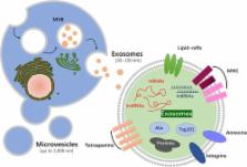
          Stem cells, with their therapeutic potential in tissue repair and regeneration, have been widely used in translational medicine. Recent evidence suggests that the beneficial effects are mediated largely by their paracrine actions rather than the engraftment and differentiation at the injured sites. Extracellular vesicles (EVs), actively released from cells, play important roles in cell‐to‐cell communication and display multiple functions in tissue regeneration. In the present report, we will briefly review the current knowledge related to the therapeutic potential of EVs, particularly stem cell or progenitor cell‐derived ones for promoting tissue repair and regeneration, and focus on the restorative properties of exosomes/microvesicles in cutaneous wound healing, bone regeneration, hindlimb ischemia, and vascular injury repair. S tem C ells T ranslational M edicine 2017;6:1753–1758

          Related collections

          Most cited references33

          • Record: found
          • Abstract: found
          • Article: found
          Is Open Access

          Exosomes released from human induced pluripotent stem cells-derived MSCs facilitate cutaneous wound healing by promoting collagen synthesis and angiogenesis

          Background Human induced pluripotent stem cell-derived mesenchymal stem cells (hiPSC-MSCs) have emerged as a promising alternative for stem cell transplantation therapy. Exosomes derived from mesenchymal stem cells (MSC-Exos) can play important roles in repairing injured tissues. However, to date, no reports have demonstrated the use of hiPSC-MSC-Exos in cutaneous wound healing, and little is known regarding their underlying mechanisms in tissue repair. Methods hiPSC-MSC-Exos were injected subcutaneously around wound sites in a rat model and the efficacy of hiPSC-MSC-Exos was assessed by measuring wound closure areas, by histological and immunofluorescence examinations. We also evaluated the in vitro effects of hiPSC-MSC-Exos on both the proliferation and migration of human dermal fibroblasts and human umbilical vein endothelial cells (HUVECs) by cell-counting and scratch assays, respectively. The effects of exosomes on fibroblast collagen and elastin secretion were studied in enzyme-linked immunosorbent assays and quantitative reverse-transcriptase–polymerase chain reaction (qRT-PCR). In vitro capillary network formation was determined in tube-formation assays. Results Transplanting hiPSC-MSC-Exos to wound sites resulted in accelerated re-epithelialization, reduced scar widths, and the promotion of collagen maturity. Moreover, hiPSC-MSC-Exos not only promoted the generation of newly formed vessels, but also accelerated their maturation in wound sites. We found that hiPSC-MSC-Exos stimulated the proliferation and migration of human dermal fibroblasts and HUVECs in a dose-dependent manner in vitro. Similarly, Type I, III collagen and elastin secretion and mRNA expression by fibroblasts and tube formation by HUVECs were also increased with increasing hiPSC-MSC-Exos concentrations. Conclusions Our findings suggest that hiPSC-MSC-Exos can facilitate cutaneous wound healing by promoting collagen synthesis and angiogenesis. These data provide the first evidence for the potential of hiPSC-MSC-Exos in treating cutaneous wounds.
            Bookmark
            • Record: found
            • Abstract: found
            • Article: not found

            Comprehensive Proteomic Analysis of Mesenchymal Stem Cell Exosomes Reveals Modulation of Angiogenesis via Nuclear Factor-KappaB Signaling.

            Mesenchymal stem cells (MSC) are known to facilitate healing of ischemic tissue related diseases through proangiogenic secretory proteins. Recent studies further show that MSC derived exosomes function as paracrine effectors of angiogenesis, however, the identity of which components of the exosome proteome responsible for this effect remains elusive. To address this we used high-resolution isoelectric focusing coupled liquid chromatography tandem mass spectrometry, an unbiased high throughput proteomics approach to comprehensively characterize the proteinaceous contents of MSCs and MSC derived exosomes. We probed the proteome of MSCs and MSC derived exosomes from cells cultured under expansion conditions and under ischemic tissue simulated conditions to elucidate key angiogenic paracrine effectors present and potentially differentially expressed in these conditions. In total, 6,342 proteins were identified in MSCs and 1,927 proteins in MSC derived exosomes, representing to our knowledge the first time these proteomes have been probed comprehensively. Multilayered analyses identified several putative paracrine effectors of angiogenesis present in MSC exosomes and increased in expression in MSCs exposed to ischemic tissue-simulated conditions; these include platelet derived growth factor, epidermal growth factor, fibroblast growth factor, and most notably nuclear factor-kappaB (NFkB) signaling pathway proteins. NFkB signaling was identified as a key mediator of MSC exosome induced angiogenesis in endothelial cells by functional in vitro validation using a specific inhibitor. Collectively, the results of our proteomic analysis show that MSC derived exosomes contain a robust profile of angiogenic paracrine effectors, which have potential for the treatment of ischemic tissue-related diseases.
              Bookmark
              • Record: found
              • Abstract: found
              • Article: not found

              Umbilical Cord-Derived Mesenchymal Stem Cell-Derived Exosomal MicroRNAs Suppress Myofibroblast Differentiation by Inhibiting the Transforming Growth Factor-β/SMAD2 Pathway During Wound Healing.

              : Excessive scar formation caused by myofibroblast aggregations is of great clinical importance during skin wound healing. Studies have shown that mesenchymal stem cells (MSCs) can promote skin regeneration, but whether MSCs contribute to scar formation remains undefined. We found that umbilical cord-derived MSCs (uMSCs) reduced scar formation and myofibroblast accumulation in a skin-defect mouse model. We found that these functions were mainly dependent on uMSC-derived exosomes (uMSC-Exos) and especially exosomal microRNAs. Through high-throughput RNA sequencing and functional analysis, we demonstrated that a group of uMSC-Exos enriched in specific microRNAs (miR-21, -23a, -125b, and -145) played key roles in suppressing myofibroblast formation by inhibiting the transforming growth factor-β2/SMAD2 pathway. Finally, using the strategy we established to block miRNAs inside the exosomes, we showed that these specific exosomal miRNAs were essential for the myofibroblast-suppressing and anti-scarring functions of uMSCs both in vitro and in vivo. Our study revealed a novel role of exosomal miRNAs in uMSC-mediated therapy, suggesting that the clinical application of uMSC-derived exosomes might represent a strategy to prevent scar formation during wound healing.
                Bookmark

                Author and article information

                Contributors
                wangyang63@126.com
                Journal
                Stem Cells Transl Med
                Stem Cells Transl Med
                10.1002/(ISSN)2157-6580
                SCT3
                Stem Cells Translational Medicine
                John Wiley and Sons Inc. (Hoboken )
                2157-6564
                2157-6580
                27 June 2017
                September 2017
                : 6
                : 9 ( doiID: 10.1002/sct3.2017.6.issue-9 )
                : 1753-1758
                Affiliations
                [ 1 ] Institute of Microsurgery on Extremities, Shanghai Jiao Tong University Affiliated Sixth People's Hospital 600 Yishan Road Shanghai 200233 People's Republic of China
                [ 2 ] Department of Orthopedic Surgery Shanghai Jiao Tong University Affiliated Sixth People's Hospital 600 Yishan Road Shanghai 200233 People's Republic of China
                Author notes
                [*] [* ]Correspondence: Yang Wang, Ph.D., Institute of Microsurgery on Extremities, Shanghai Jiao Tong University Affiliated Sixth People's Hospital, Shanghai 200233, China. Telephone: 86‐21‐24056319; e‐mail: wangyang63@ 123456126.com
                Author information
                http://orcid.org/0000-0002-6649-5291
                Article
                SCT312172
                10.1002/sctm.16-0477
                5689748
                28653443
                29b6458e-4a52-4110-b892-6f136d08dce9
                © 2017 The Authors S tem C ells T ranslational M edicine published by Wiley Periodicals, Inc. on behalf of AlphaMed Press

                This is an open access article under the terms of the Creative Commons Attribution‐NonCommercial‐NoDerivs License, which permits use and distribution in any medium, provided the original work is properly cited, the use is non‐commercial and no modifications or adaptations are made.

                History
                : 29 November 2016
                : 21 April 2017
                Page count
                Figures: 2, Tables: 3, Pages: 7, Words: 5223
                Categories
                Perspectives
                Bone Marrow Stem Cells
                Cord / Cord Blood Stem Cells (WJ‐MSCs)
                Induced Pluripotent stem cells
                Mesenchymal Stem Cells
                Angiogenesis
                Osteogenesis
                Wound Healing / Fibrosis
                Vascular Disease
                Perspectives
                Perspectives
                Custom metadata
                2.0
                sct312172
                September 2017
                Converter:WILEY_ML3GV2_TO_NLMPMC version:5.2.4.1 mode:remove_FC converted:09.11.2017

                Comments

                Comment on this article