34
views
0
recommends
+1 Recommend
0 collections
    0
    shares
      • Record: found
      • Abstract: found
      • Article: found
      Is Open Access

      Activation of mitochondrial TUFM ameliorates metabolic dysregulation through coordinating autophagy induction

      research-article

      Read this article at

      Bookmark
          There is no author summary for this article yet. Authors can add summaries to their articles on ScienceOpen to make them more accessible to a non-specialist audience.

          Abstract

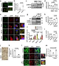
          Disorders of autophagy, a key regulator of cellular homeostasis, cause a number of human diseases. Due to the role of autophagy in metabolic dysregulation, there is a need to identify autophagy regulators as therapeutic targets. To address this need, we conducted an autophagy phenotype-based screen and identified the natural compound kaempferide (Kaem) as an autophagy enhancer. Kaem promoted autophagy through translocation of transcription factor EB (TFEB) without MTOR perturbation, suggesting it is safe for administration. Moreover, Kaem accelerated lipid droplet degradation in a lysosomal activity-dependent manner in vitro and ameliorated metabolic dysregulation in a diet-induced obesity mouse model. To elucidate the mechanism underlying Kaem’s biological activity, the target protein was identified via combined drug affinity responsive target stability and LC–MS/MS analyses. Kaem directly interacted with the mitochondrial elongation factor TUFM, and TUFM absence reversed Kaem-induced autophagy and lipid degradation. Kaem also induced mitochondrial reactive oxygen species (mtROS) to sequentially promote lysosomal Ca 2+ efflux, TFEB translocation and autophagy induction, suggesting a role of TUFM in mtROS regulation. Collectively, these results demonstrate that Kaem is a potential therapeutic candidate/chemical tool for treating metabolic dysregulation and reveal a role for TUFM in autophagy for metabolic regulation with lipid overload.

          Abstract

          Kim, Hwang et al. use in vitro and in vivo models of autophagy disorder/metabolic dysfunction to show that in this context, the natural compound kaempferide is an autophagy enhancer and reveal that one of the underlying mechanisms governing this is mediated by the mitochondrial elongation factor TUFM. This insight may have therapeutic value in the treatment of metabolic disorders.

          Related collections

          Most cited references63

          • Record: found
          • Abstract: found
          • Article: not found

          The coming of age of chaperone-mediated autophagy

          Chaperone-mediated autophagy (CMA) was the first studied process that indicated that degradation of intracellular components by the lysosome can be selective — a concept that is now well accepted for other forms of autophagy. Lysosomes can degrade cellular cytosol in a nonspecific manner but can also discriminate what to target for degradation with the involvement of a degradation tag, a chaperone and a sophisticated mechanism to make the selected proteins cross the lysosomal membrane through a dedicated translocation complex. Recent studies modulating CMA activity in vivo using transgenic mouse models have demonstrated that selectivity confers on CMA the ability to participate in the regulation of multiple cellular functions. Timely degradation of specific cellular proteins by CMA modulates, for example, glucose and lipid metabolism, DNA repair, cellular reprograming and the cellular response to stress. These findings expand the physiological relevance of CMA beyond its originally identified role in protein quality control and reveal that CMA failure with age may aggravate diseases, such as ageing-associated neurodegeneration and cancer.
            Bookmark
            • Record: found
            • Abstract: found
            • Article: found
            Is Open Access

            Mitochondrial electron transport chain, ROS generation and uncoupling (Review)

            The mammalian mitochondrial electron transport chain (ETC) includes complexes I-IV, as well as the electron transporters ubiquinone and cytochrome c. There are two electron transport pathways in the ETC: Complex I/III/IV, with NADH as the substrate and complex II/III/IV, with succinic acid as the substrate. The electron flow is coupled with the generation of a proton gradient across the inner membrane and the energy accumulated in the proton gradient is used by complex V (ATP synthase) to produce ATP. The first part of this review briefly introduces the structure and function of complexes I-IV and ATP synthase, including the specific electron transfer process in each complex. Some electrons are directly transferred to O2 to generate reactive oxygen species (ROS) in the ETC. The second part of this review discusses the sites of ROS generation in each ETC complex, including sites IF and IQ in complex I, site IIF in complex II and site IIIQo in complex III, and the physiological and pathological regulation of ROS. As signaling molecules, ROS play an important role in cell proliferation, hypoxia adaptation and cell fate determination, but excessive ROS can cause irreversible cell damage and even cell death. The occurrence and development of a number of diseases are closely related to ROS overproduction. Finally, proton leak and uncoupling proteins (UCPS) are discussed. Proton leak consists of basal proton leak and induced proton leak. Induced proton leak is precisely regulated and induced by UCPs. A total of five UCPs (UCP1-5) have been identified in mammalian cells. UCP1 mainly plays a role in the maintenance of body temperature in a cold environment through non-shivering thermogenesis. The core role of UCP2-5 is to reduce oxidative stress under certain conditions, therefore exerting cytoprotective effects. All diseases involving oxidative stress are associated with UCPs.
              Bookmark
              • Record: found
              • Abstract: found
              • Article: not found

              Lysosomal calcium signaling regulates autophagy via calcineurin and TFEB

              The view of the lysosome as the terminal end of cellular catabolic pathways has been challenged by recent studies showing a central role of this organelle in the control of cell function. Here we show that a lysosomal Ca2+ signaling mechanism controls the activities of the phosphatase calcineurin and of its substrate TFEB, a master transcriptional regulator of lysosomal biogenesis and autophagy. Lysosomal Ca2+ release via mucolipin 1 (MCOLN1) activates calcineurin, which binds and de-phosphorylates TFEB, thus promoting its nuclear translocation. Genetic and pharmacological inhibition of calcineurin suppressed TFEB activity during starvation and physical exercise, while calcineurin overexpression and constitutive activation had the opposite effect. Induction of autophagy and lysosomal biogenesis via TFEB required MCOLN1-mediated calcineurin activation, linking lysosomal calcium signaling to both calcineurin regulation and autophagy induction. Thus, the lysosome reveals itself as a hub for the signaling pathways that regulate cellular homeostasis.
                Bookmark

                Author and article information

                Contributors
                kwonhj@yonsei.ac.kr
                Journal
                Commun Biol
                Commun Biol
                Communications Biology
                Nature Publishing Group UK (London )
                2399-3642
                4 January 2021
                4 January 2021
                2021
                : 4
                : 1
                Affiliations
                [1 ]GRID grid.15444.30, ISNI 0000 0004 0470 5454, Chemical Genomics Global Research Laboratory, Department of Biotechnology, College of Life Science and Biotechnology, , Yonsei University, ; Seoul, 03722 Republic of Korea
                [2 ]GRID grid.410885.0, ISNI 0000 0000 9149 5707, Biomedical Omics Group, , Korea Basic Science Institute, ; Ochang, Chungbuk 28119 Republic of Korea
                Author information
                http://orcid.org/0000-0003-1314-2940
                http://orcid.org/0000-0001-9223-1428
                http://orcid.org/0000-0002-6919-833X
                Article
                1566
                10.1038/s42003-020-01566-0
                7782552
                33398033
                2e84a3dd-3663-4b0d-ac8b-51f5100eb4ac
                © The Author(s) 2021

                Open Access This article is licensed under a Creative Commons Attribution 4.0 International License, which permits use, sharing, adaptation, distribution and reproduction in any medium or format, as long as you give appropriate credit to the original author(s) and the source, provide a link to the Creative Commons license, and indicate if changes were made. The images or other third party material in this article are included in the article’s Creative Commons license, unless indicated otherwise in a credit line to the material. If material is not included in the article’s Creative Commons license and your intended use is not permitted by statutory regulation or exceeds the permitted use, you will need to obtain permission directly from the copyright holder. To view a copy of this license, visit http://creativecommons.org/licenses/by/4.0/.

                History
                : 16 April 2020
                : 2 December 2020
                Categories
                Article
                Custom metadata
                © The Author(s) 2021

                molecular medicine,mechanism of action
                molecular medicine, mechanism of action

                Comments

                Comment on this article