58
views
0
recommends
+1 Recommend
1 collections
    0
    shares
      • Record: found
      • Abstract: found
      • Article: found
      Is Open Access

      Structure of cyclin G-associated kinase (GAK) trapped in different conformations using nanobodies

      research-article
      * , * , , , § , § , , , , , , , § , * , * , 2
      Biochemical Journal
      Portland Press Ltd.
      activation loop, cyclin G-associated kinase, drug side effect, kinase inhibitor, nanobody, protein structure, ASCH, activation segment C-terminal helix, AUC, analytical ultracentrifugation, CDR, complementarity-determining region, DARPin, designed ankyrin-repeat protein, EGFR, epidermal growth factor receptor, GAK, cyclin G-associated kinase, HA, haemagglutinin, MPSK1, myristoylated and palmitoylated serine/threonine kinase 1, NAK, numb-associated kinase, Nb, nanobody, RU, resonance unit, SeMet, selenomethionine, SPR, surface plasmon resonance, TCEP, tris-(2-carboxyethyl)phosphine, TEV, Tobacco etch virus

      Read this article at

      Bookmark
          There is no author summary for this article yet. Authors can add summaries to their articles on ScienceOpen to make them more accessible to a non-specialist audience.

          Abstract

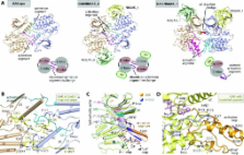
          GAK (cyclin G-associated kinase) is a key regulator of clathrin-coated vesicle trafficking and plays a central role during development. Additionally, due to the unusually high plasticity of its catalytic domain, it is a frequent ‘off-target’ of clinical kinase inhibitors associated with respiratory side effects of these drugs. In the present paper, we determined the crystal structure of the GAK catalytic domain alone and in complex with specific single-chain antibodies (nanobodies). GAK is constitutively active and weakly associates in solution. The GAK apo structure revealed a dimeric inactive state of the catalytic domain mediated by an unusual activation segment interaction. Co-crystallization with the nanobody NbGAK_4 trapped GAK in a dimeric arrangement similar to the one observed in the apo structure, whereas NbGAK_1 captured the activation segment of monomeric GAK in a well-ordered conformation, representing features of the active kinase. The presented structural and biochemical data provide insight into the domain plasticity of GAK and demonstrate the utility of nanobodies to gain insight into conformational changes of dynamic molecules. In addition, we present structural data on the binding mode of ATP mimetic inhibitors and enzyme kinetic data, which will support rational inhibitor design of inhibitors to reduce the off-target effect on GAK.

          Abstract

          Cyclin G-associated kinase (GAK) is a regulator of clathrin-coated vesicle trafficking. The determined crystal structures of GAK in complex with specific single chain antibodies (nanobodies) revealed the domain plasticity of this kinase and unusual activation segment architecture.

          Related collections

          Most cited references33

          • Record: found
          • Abstract: found
          • Article: not found

          The protein kinase complement of the human genome.

          G. Manning (2002)
          We have catalogued the protein kinase complement of the human genome (the "kinome") using public and proprietary genomic, complementary DNA, and expressed sequence tag (EST) sequences. This provides a starting point for comprehensive analysis of protein phosphorylation in normal and disease states, as well as a detailed view of the current state of human genome analysis through a focus on one large gene family. We identify 518 putative protein kinase genes, of which 71 have not previously been reported or described as kinases, and we extend or correct the protein sequences of 56 more kinases. New genes include members of well-studied families as well as previously unidentified families, some of which are conserved in model organisms. Classification and comparison with model organism kinomes identified orthologous groups and highlighted expansions specific to human and other lineages. We also identified 106 protein kinase pseudogenes. Chromosomal mapping revealed several small clusters of kinase genes and revealed that 244 kinases map to disease loci or cancer amplicons.
            Bookmark
            • Record: found
            • Abstract: found
            • Article: not found

            Protein kinases: evolution of dynamic regulatory proteins.

            Eukayotic protein kinases evolved as a family of highly dynamic molecules with strictly organized internal architecture. A single hydrophobic F-helix serves as a central scaffold for assembly of the entire molecule. Two non-consecutive hydrophobic structures termed "spines" anchor all the elements important for catalysis to the F-helix. They make firm, but flexible, connections within the molecule, providing a high level of internal dynamics of the protein kinase. During the course of evolution, protein kinases developed a universal regulatory mechanism associated with a large activation segment that can be dynamically folded and unfolded in the course of cell functioning. Protein kinases thus represent a unique, highly dynamic, and precisely regulated set of switches that control most biological events in eukaryotic cells. Copyright © 2010. Published by Elsevier Ltd.
              Bookmark
              • Record: found
              • Abstract: found
              • Article: not found

              Surface comparison of active and inactive protein kinases identifies a conserved activation mechanism.

              The surface comparison of different serine-threonine and tyrosine kinases reveals a set of 30 residues whose spatial positions are highly conserved. The comparison between active and inactive conformations identified the residues whose positions are the most sensitive to activation. Based on these results, we propose a model of protein kinase activation. This model explains how the presence of a phosphate group in the activation loop determines the position of the catalytically important aspartate in the Asp-Phe-Gly motif. According to the model, the most important feature of the activation is a "spine" formation that is dynamically assembled in all active kinases. The spine is comprised of four hydrophobic residues that we detected in a set of 23 eukaryotic and prokaryotic kinases. It spans the molecule and plays a coordinating role in activated kinases. The spine is disordered in the inactive kinases and can explain how stabilization of the whole molecule is achieved upon phosphorylation.
                Bookmark

                Author and article information

                Journal
                Biochem J
                Biochem. J
                bic
                BJ
                Biochemical Journal
                Portland Press Ltd.
                0264-6021
                1470-8728
                17 January 2014
                14 March 2014
                1 April 2014
                : 459
                : Pt 1
                : 59-69
                Affiliations
                *University of Oxford, Target Discovery Institute (TDI) and Structural Genomics Consortium (SGC), Old Road Campus Research Building, Oxford OX3 7DQ, U.K.
                †Research Unit of Cellular and Molecular Immunology and Department of Structural Biology, VIB, Vrije Universiteit Brussel, Pleinlaan 2, 1050 Brussels, Belgium
                ‡Department of Biochemistry, University of Kassel, Heinrich-Plett Strasse 40, 34132 Kassel, Germany
                §Biaffin GmbH & CoKG, Heinrich-Plett Strasse 40, 34132 Kassel, Germany
                ¶Department of Animal Medicine and Surgery, Veterinary Faculty, University of Las Palmas de Gran Canaria, 35416, Arucas, Las Palmas, Spain
                ∥Midwest Center for Structural Genomics and Structural Biology Center, Biosciences Division, Argonne National Laboratory, 9700 South Cass Avenue, Argonne, IL 60439, U.S.A.
                Author notes

                1These authors contributed equally to the work.

                2To whom correspondence should be addressed (email susanne.muller-knapp@ 123456sgc.ox.ac.uk ).

                The crystal structures reported in this paper have been deposited in the PDB under codes 4O38, 4C57, 4C58 and 4C59.

                Article
                BJ20131399
                10.1042/BJ20131399
                3957475
                24438162
                2eb206ea-fa7d-4313-bbe1-bcb6835e982c
                © 2014 The author(s) has paid for this article to be freely available under the terms of the Creative Commons Attribution Licence (CC-BY)(http://creativecommons.org/licenses/by/3.0/) which permits unrestricted use, distribution and reproduction in any medium, provided the original work is properly cited.

                This is an open-access article distributed under the terms of the Creative Commons Attribution License, which permits unrestricted use, distribution and reproduction in any medium, provided the original work is properly cited.

                History
                : 22 October 2013
                : 16 January 2014
                : 17 January 2014
                Page count
                Figures: 4, Tables: 2, References: 49, Pages: 11
                Categories
                Research Article

                Biochemistry
                activation loop,cyclin g-associated kinase,drug side effect,kinase inhibitor,nanobody,protein structure,asch, activation segment c-terminal helix,auc, analytical ultracentrifugation,cdr, complementarity-determining region,darpin, designed ankyrin-repeat protein,egfr, epidermal growth factor receptor,gak, cyclin g-associated kinase,ha, haemagglutinin,mpsk1, myristoylated and palmitoylated serine/threonine kinase 1,nak, numb-associated kinase,nb, nanobody,ru, resonance unit,semet, selenomethionine,spr, surface plasmon resonance,tcep, tris-(2-carboxyethyl)phosphine,tev, tobacco etch virus

                Comments

                Comment on this article