1
views
0
recommends
+1 Recommend
1 collections
    0
    shares
      • Record: found
      • Abstract: found
      • Article: found
      Is Open Access

      Alteration of Diffusion Capacity After SARS-CoV-2 Infection: A Pathophysiological Approach

      research-article

      Read this article at

      Bookmark
          There is no author summary for this article yet. Authors can add summaries to their articles on ScienceOpen to make them more accessible to a non-specialist audience.

          Abstract

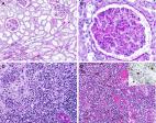
          Severe acute respiratory syndrome coronavirus 2 ( SARS- CoV- 2) infection has affected millions of people worldwide, and pneumonia affects 90% of patients. This raises the possibility of millions of people with altered lung function. Few data exist to date on pulmonary function after SARS-CoV-2 infection, but alteration of diffusion capacity of CO ( D LCO) is the most frequently described abnormality. First, we present original data on lung function at 3 months after SARS-CoV-2 infection and discuss the effect of using European Coal and Steel Community (ECSC) or Global Lung Function Initiative (GLI) reference equations to diagnose diffusion capacity. Second, we review existing data on D LCO alteration after SARS-CoV-2 infection and discuss the implication of restrictive disorder in D LCO alteration. Last, we discuss the pathophysiology of D LCO alteration and try to disentangle vascular damage and fibrosis.

          Related collections

          Most cited references48

          • Record: found
          • Abstract: found
          • Article: not found

          Tissue distribution of ACE2 protein, the functional receptor for SARS coronavirus. A first step in understanding SARS pathogenesis

          Abstract Severe acute respiratory syndrome (SARS) is an acute infectious disease that spreads mainly via the respiratory route. A distinct coronavirus (SARS‐CoV) has been identified as the aetiological agent of SARS. Recently, a metallopeptidase named angiotensin‐converting enzyme 2 (ACE2) has been identified as the functional receptor for SARS‐CoV. Although ACE2 mRNA is known to be present in virtually all organs, its protein expression is largely unknown. Since identifying the possible route of infection has major implications for understanding the pathogenesis and future treatment strategies for SARS, the present study investigated the localization of ACE2 protein in various human organs (oral and nasal mucosa, nasopharynx, lung, stomach, small intestine, colon, skin, lymph nodes, thymus, bone marrow, spleen, liver, kidney, and brain). The most remarkable finding was the surface expression of ACE2 protein on lung alveolar epithelial cells and enterocytes of the small intestine. Furthermore, ACE2 was present in arterial and venous endothelial cells and arterial smooth muscle cells in all organs studied. In conclusion, ACE2 is abundantly present in humans in the epithelia of the lung and small intestine, which might provide possible routes of entry for the SARS‐CoV. This epithelial expression, together with the presence of ACE2 in vascular endothelium, also provides a first step in understanding the pathogenesis of the main SARS disease manifestations. Copyright © 2004 Pathological Society of Great Britain and Ireland. Published by John Wiley & Sons, Ltd.
            Bookmark
            • Record: found
            • Abstract: found
            • Article: not found

            Complement associated microvascular injury and thrombosis in the pathogenesis of severe COVID-19 infection: A report of five cases

            Acute respiratory failure and a systemic coagulopathy are critical aspects of the morbidity and mortality characterizing infection with severe acute respiratory distress syndrome-associated coronavirus-2 (SARS-CoV-2), the etiologic agent of Coronavirus disease 2019 (COVID-19). We examined skin and lung tissues from 5 patients with severe COVID-19 characterized by respiratory failure (n=5) and purpuric skin rash (n=3). The pattern of COVID-19 pneumonitis was predominantly a pauci-inflammatory septal capillary injury with significant septal capillary mural and luminal fibrin deposition and permeation of the inter-alveolar septa by neutrophils. No viral cytopathic changes were observed and the diffuse alveolar damage (DAD) with hyaline membranes, inflammation, and type II pneumocyte hyperplasia, hallmarks of classic ARDS, were not prominent. These pulmonary findings were accompanied by significant deposits of terminal complement components C5b-9 (membrane attack complex), C4d, and mannose binding lectin (MBL)-associated serine protease (MASP)2, in the microvasculature, consistent with sustained, systemic activation of the alternative and lectin-based complement pathways. The purpuric skin lesions similarly showed a pauci-inflammatory thrombogenic vasculopathy, with deposition of C5b-9 and C4d in both grossly involved and normally-appearing skin. In addition, there was co-localization of COVID-19 spike glycoproteins with C4d and C5b-9 in the inter-alveolar septa and the cutaneous microvasculature of two cases examined. In conclusion, at least a subset of sustained, severe COVID-19 may define a type of catastrophic microvascular injury syndrome mediated by activation of complement pathways and an associated procoagulant state. It provides a foundation for further exploration of the pathophysiologic importance of complement in COVID-19, and could suggest targets for specific intervention.
              Bookmark
              • Record: found
              • Abstract: found
              • Article: found
              Is Open Access

              Postmortem examination of COVID‐19 patients reveals diffuse alveolar damage with severe capillary congestion and variegated findings in lungs and other organs suggesting vascular dysfunction

              Aims Coronavirus disease 2019 (COVID‐19), caused by severe acute respiratory syndrome coronavirus 2 (SARS‐CoV‐2), has rapidly evolved into a sweeping pandemic. Its major manifestation is in the respiratory tract, and the general extent of organ involvement and the microscopic changes in the lungs remain insufficiently characterised. Autopsies are essential to elucidate COVID‐19‐associated organ alterations. Methods and results This article reports the autopsy findings of 21 COVID‐19 patients hospitalised at the University Hospital Basel and at the Cantonal Hospital Baselland, Switzerland. An in‐corpore technique was performed to ensure optimal staff safety. The primary cause of death was respiratory failure with exudative diffuse alveolar damage and massive capillary congestion, often accompanied by microthrombi despite anticoagulation. Ten cases showed superimposed bronchopneumonia. Further findings included pulmonary embolism (n = 4), alveolar haemorrhage (n = 3), and vasculitis (n = 1). Pathologies in other organ systems were predominantly attributable to shock; three patients showed signs of generalised and five of pulmonary thrombotic microangiopathy. Six patients were diagnosed with senile cardiac amyloidosis upon autopsy. Most patients suffered from one or more comorbidities (hypertension, obesity, cardiovascular diseases, and diabetes mellitus). Additionally, there was an overall predominance of males and individuals with blood group A (81% and 65%, respectively). All relevant histological slides are linked as open‐source scans in supplementary files. Conclusions This study provides an overview of postmortem findings in COVID‐19 cases, implying that hypertensive, elderly, obese, male individuals with severe cardiovascular comorbidities as well as those with blood group A may have a lower threshold of tolerance for COVID‐19. This provides a pathophysiological explanation for higher mortality rates among these patients.
                Bookmark

                Author and article information

                Contributors
                Journal
                Front Physiol
                Front Physiol
                Front. Physiol.
                Frontiers in Physiology
                Frontiers Media S.A.
                1664-042X
                25 March 2021
                2021
                25 March 2021
                : 12
                : 624062
                Affiliations
                [1] 1Neurodiderot, INSERM, Université de Paris , Paris, France
                [2] 2Physiologie-Explorations Fonctionnelles, FHU APOLLO, Assistance Publique Hôpitaux de Paris, Hôpital Bichat-Claude Bernard , Paris, France
                [3] 3Centre de Ressources et de Compétences de la Mucoviscidose, Service de Pneumologie et Explorations Fonctionnelles Respiratoires, CHRU de Tours , Tours, France
                [4] 4CEPR/INSERM, UMR 1100, Université de Tours , Tours, France
                [5] 5Laboratoire Matière et Systèmes Complexes, UMR 7505, CNRS , Paris, France
                [6] 6INSERM U 1149, Center for Research in Inflammation, Université de Paris , Paris, France
                Author notes

                Edited by: Ravi Nistala, University of Missouri, United States

                Reviewed by: Jordan A. Guenette, University of British Columbia, Canada; Hariharan Regunath, University of Missouri, United States

                *Correspondence: Justine Frija-Masson, justine.frija@ 123456aphp.fr ; orcid.org/0000-0001-5575-3913

                This article was submitted to Respiratory Physiology, a section of the journal Frontiers in Physiology

                Article
                10.3389/fphys.2021.624062
                8030254
                33841169
                3262bb4f-2e99-4868-b3c2-ccedbb8b4543
                Copyright © 2021 Frija-Masson, Bancal, Plantier, Benzaquen, Mangin, Penaud, Arnoult, Flamant and d’Ortho.

                This is an open-access article distributed under the terms of the Creative Commons Attribution License (CC BY). The use, distribution or reproduction in other forums is permitted, provided the original author(s) and the copyright owner(s) are credited and that the original publication in this journal is cited, in accordance with accepted academic practice. No use, distribution or reproduction is permitted which does not comply with these terms.

                History
                : 30 October 2020
                : 27 January 2021
                Page count
                Figures: 1, Tables: 1, Equations: 0, References: 50, Pages: 7, Words: 0
                Categories
                Physiology
                Perspective

                Anatomy & Physiology
                sars-cov-2,dlco,pneumonia,pulmonary function test,covid-19
                Anatomy & Physiology
                sars-cov-2, dlco, pneumonia, pulmonary function test, covid-19

                Comments

                Comment on this article