82
views
0
recommends
+1 Recommend
0 collections
    0
    shares
      • Record: found
      • Abstract: found
      • Article: found
      Is Open Access

      Development of New Cardiac Deformity Indexes for Pectus Excavatum on Computed Tomography: Feasibility for Pre- and Post-Operative Evaluation

      research-article

      Read this article at

      Bookmark
          There is no author summary for this article yet. Authors can add summaries to their articles on ScienceOpen to make them more accessible to a non-specialist audience.

          Abstract

          Purpose

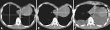
          The aim of this study was to evaluate new cardiac deformity indexes (CDIs) for diagnosis of pectus excavatum as well as morphological assessment of heart on computed tomography (CT).

          Materials and Methods

          We retrospectively evaluated the CT images of the control group (n=200), and the pectus excavatum before and after correction groups (n=178), and calculated the CDIs; cardiac compression index (CCI), and cardiac asymmetry index (CAI). We also calculated chest wall compression index (CWCI) and asymmetry index (CWAI) on the axial images. We performed logistic regression analysis using each index and age as predictor variables.

          Results

          The CDIs (CCI and CAI) were significant ( p < 0.05) in the diagnosis of pectus excavatum, regardless of age ( p = 0.4033, p = 0.8113). The CWCI and CWAI were significant ( p < 0.05) and significantly affected by age ( p < 0.05). If we selected 1.82 as the cutoff of the CCI, the sensitivity and specificity were 99.4% and 98%, respectively. The following cutoffs and the sensitivity and specificity were obtained: 1.15 for the CAI gave 94.4% and 94.5%, 3.05 for the CWCI gave 92.1% and 92%, and 1 for the CWAI gave 62.4% and 65%, respectively. The CCI after repair improved from 2.83 ± 0.84 to 1.84 ± 0.33, while the CWCI improved from 4.49 ± 1.61 to 2.57 ± 0.44.

          Conclusion

          CDIs such as the CCI and CAI may be potentially useful to detect and estimate repair for pectus excavatum.

          Related collections

          Most cited references13

          • Record: found
          • Abstract: found
          • Article: not found

          A 10-year review of a minimally invasive technique for the correction of pectus excavatum.

          The aim of this study was to assess the results of a 10-year experience with a minimally invasive operation that requires neither cartilage incision nor resection for correction of pectus excavatum. From 1987 to 1996, 148 patients were evaluated for chest wall deformity. Fifty of 127 patients suffering from pectus excavatum were selected for surgical correction. Eight older patients underwent the Ravitch procedure, and 42 patients under age 15 were treated by the minimally invasive technique. A convex steel bar is inserted under the sternum through small bilateral thoracic incisions. The steel bar is inserted with the convexity facing posteriorly, and when it is in position, the bar is turned over, thereby correcting the deformity. After 2 years, when permanent remolding has occurred, the bar is removed in an outpatient procedure. Of 42 patients who had the minimally invasive procedure, 30 have undergone bar removal. Initial excellent results were maintained in 22, good results in four, fair in two, and poor in two, with mean follow-up since surgery of 4.6 years (range, 1 to 9.2 years). Mean follow-up since bar removal is 2.8 years (range, 6 months to 7 years). Average blood loss was 15 mL. Average length of hospital stay was 4.3 days. Patients returned to full activity after 1 month. Complications were pneumothorax in four patients, requiring thoracostomy in one patient; superficial wound infection in one patient; and displacement of the steel bar requiring revision in two patients. The fair and poor results occurred early in the series because (1) the bar was too soft (three patients), (2) the sternum was too soft in one of the patients with Marfan's syndrome, and (3) in one patient with complex thoracic anomalies, the bar was removed too soon. This minimally invasive technique, which requires neither cartilage incision nor resection, is effective. Since increasing the strength of the steel bar and inserting two bars where necessary, we have had excellent long-term results. The upper limits of age for this procedure require further evaluation.
            Bookmark
            • Record: found
            • Abstract: found
            • Article: not found

            Use of CT scans in selection of patients for pectus excavatum surgery: a preliminary report.

            A pectus index can be derived from dividing the transverse diameter of the chest by the anterior-posterior diameter on a simple CT scan. In a preliminary report, all patients who required operative correction for pectus excavatum had a pectus index greater than 3.25 while matched normal controls were all less than 3.25. A simple CT scan may be a useful adjunct in objective evaluation of children and teenagers for surgery of pectus excavatum.
              Bookmark
              • Record: found
              • Abstract: found
              • Article: not found

              The Nuss procedure for pectus excavatum: evolution of techniques and early results on 322 patients.

              The Nuss procedure is a minimally invasive technique using a retrosternal bar to repair pectus excavatum. Although its technical simplicity and cosmetic advantages are remarkable, early applications have been limited to children with symmetrical pectus excavatum. We report a large single-institution experience including technical modifications to correct asymmetric configurations and extend the procedure to adult patients. We retrospectively reviewed 322 consecutive patients who underwent repair of pectus excavatum by the Nuss technique and its modifications between August 1999 and June 2002. Of the patients 251 (78%) were children and 71 (22%) were adults. Precise morphologic characterization of the pectus allowed appropriate shaping of the bar to achieve a symmetric repair. Of the 322, 185 (57%) had symmetric and 137 (43%) had asymmetric pectus excavatum. Within the asymmetric group 71 were eccentric, 47 were unbalanced, and 19 were combined. Modifications to the shape of the bar including asymmetric and seagull bars were developed to deal with these types of asymmetry. A double bar or compound bar technique was used in most of the adults. Multipoint wire fixations to ribs were utilized to prevent bar rotation. Postoperative complications included pneumothorax (n = 24, 7.5%) and bar displacement (n = 11, 3.4%). The bar was removed in 42 patients 2 years after the initial procedure. Precise morphologic classification has led to modifications of the Nuss technique that facilitate correction of virtually all varieties of pectus excavatum including patients with asymmetric varieties and adults.
                Bookmark

                Author and article information

                Journal
                Yonsei Med J
                YMJ
                Yonsei Medical Journal
                Yonsei University College of Medicine
                0513-5796
                1976-2437
                30 June 2009
                23 June 2009
                : 50
                : 3
                : 385-390
                Affiliations
                [1 ]Department of Diagnostic Radiology, Korea University Hospital, Ansan, Korea.
                [2 ]Department of Thoracic Surgery, Korea University Hospital, Ansan, Korea.
                [3 ]Department of Biostatistics, Korea University Hospital, Ansan, Korea.
                Author notes
                Corresponding author: Dr. Ki Yeol Lee, Department of Diagnostic Radiology, Korea University Hospital, 516 Gojan-dong, Ansan 425-707, Korea. Tel: 81-31-412-5227, Fax: 81-31-412-5224, kiylee@ 123456korea.ac.kr
                Article
                10.3349/ymj.2009.50.3.385
                2703762
                19568601
                33df7fa6-e0f5-450a-93c7-9108a44e4e7b
                © Copyright: Yonsei University College of Medicine 2009

                This is an Open Access article distributed under the terms of the Creative Commons Attribution Non-Commercial License ( http://creativecommons.org/licenses/by-nc/3.0) which permits unrestricted noncommercial use, distribution, and reproduction in any medium, provided the original work is properly cited.

                History
                : 25 August 2008
                : 29 October 2008
                : 29 October 2008
                Categories
                Original Article

                Medicine
                minimally invasive repair of pectus excavatum (mirpe),cardiac deformity index,ct scan,pectus excavatum

                Comments

                Comment on this article