7
views
0
recommends
+1 Recommend
0 collections
    0
    shares
      • Record: found
      • Abstract: found
      • Article: found
      Is Open Access

      Slower alpha rhythm associates with poorer seizure control in epilepsy

      research-article

      Read this article at

      Bookmark
          There is no author summary for this article yet. Authors can add summaries to their articles on ScienceOpen to make them more accessible to a non-specialist audience.

          Abstract

          Objective

          Slowing and frontal spread of the alpha rhythm have been reported in multiple epilepsy syndromes. We investigated whether these phenomena are associated with seizure control.

          Methods

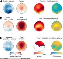
          We prospectively acquired resting‐state electroencephalogram ( EEG) in 63 patients with focal and idiopathic generalized epilepsy ( FE and IGE) and 39 age‐ and gender‐matched healthy subjects (HS). Patients were divided into good and poor (≥4 seizures/12 months) seizure control groups based on self‐reports and clinical records. We computed spectral power from 20‐sec EEG segments during eyes‐closed wakefulness, free of interictal abnormalities, and quantified power in high‐ and low‐alpha bands. Analysis of covariance and post hoc t‐tests were used to assess group differences in alpha‐power shift across all EEG channels. Permutation‐based statistics were used to assess the topography of this shift across the whole scalp.

          Results

          Compared to HS, patients showed a statistically significant shift of spectral power from high‐ to low‐alpha frequencies (effect size g = 0.78 [95% confidence interval 0.43, 1.20]). This alpha‐power shift was driven by patients with poor seizure control in both FE and IGE ( g = 1.14, [0.65, 1.74]), and occurred over midline frontal and bilateral occipital regions. IGE exhibited less alpha power shift compared to FE over bilateral frontal regions ( g = −1.16 [−0.68, −1.74]). There was no interaction between syndrome and seizure control. Effects were independent of antiepileptic drug load, time of day, or subgroup definitions.

          Interpretation

          Alpha slowing and anteriorization are a robust finding in patients with epilepsy and might represent a generic indicator of seizure liability.

          Related collections

          Most cited references26

          • Record: found
          • Abstract: found
          • Article: found
          Is Open Access

          Structural brain abnormalities in the common epilepsies assessed in a worldwide ENIGMA study

          Structural MRI abnormalities are inconsistently reported in epilepsy. In the largest neuroimaging study to date, Whelan et al. report robust structural alterations across and within epilepsy syndromes, including shared volume loss in the thalamus, and widespread cortical thickness differences. The resulting neuroanatomical map will guide prospective studies of disease progression.
            Bookmark
            • Record: found
            • Abstract: found
            • Article: not found

            Computation of measures of effect size for neuroscience data sets.

            The overwhelming majority of research in the neurosciences employs P-values stemming from tests of statistical significance to decide on the presence or absence of an effect of some treatment variable. Although a continuous variable, the P-value is commonly used to reach a dichotomous decision about the presence of an effect around an arbitrary criterion of 0.05. This analysis strategy is widely used, but has been heavily criticized in the past decades. To counter frequent misinterpretations of P-values, it has been advocated to complement or replace P-values with measures of effect size (MES). Many psychological, biological and medical journals now recommend reporting appropriate MES. One hindrance to the more frequent use of MES may be their scarcity in standard statistical software packages. Also, the arguably most widespread data analysis software in neuroscience, matlab, does not provide MES beyond correlation and receiver-operating characteristic analysis. Here we review the most common criticisms of significance testing and provide several examples from neuroscience where use of MES conveys insights not amenable through the use of P-values alone. We introduce an open-access matlab toolbox providing a wide range of MES to complement the frequently used types of hypothesis tests, such as t-tests and analysis of variance. The accompanying documentation provides calculation formulae, intuitive explanations and example calculations for each measure. The toolbox described is usable without sophisticated statistical knowledge and should be useful to neuroscientists wishing to enhance their repertoire of statistical reporting. © 2011 The Authors. European Journal of Neuroscience © 2011 Federation of European Neuroscience Societies and Blackwell Publishing Ltd.
              Bookmark
              • Record: found
              • Abstract: found
              • Article: not found

              Scalp electrical recording during paralysis: quantitative evidence that EEG frequencies above 20 Hz are contaminated by EMG.

              To identify the possible contribution of electromyogram (EMG) to scalp electroencephalogram (EEG) rhythms at rest and induced or evoked by cognitive tasks. Scalp EEG recordings were made on two subjects in presence and absence of complete neuromuscular blockade, sparing the dominant arm. The subjects undertook cognitive tasks in both states to allow direct comparison of electrical recordings. EEG rhythms in the paralysed state differed significantly compared with the unparalysed state, with 10- to 200-fold differences in the power of frequencies above 20 Hz during paralysis. Most of the scalp EEG recording above 20 Hz is of EMG origin. Previous studies measuring gamma EEG need to be re-evaluated. This has a significant impact on measurements of gamma rhythms from the scalp EEG in unparalysed humans. It is to be hoped that signal separation methods will be able to rectify this situation.
                Bookmark

                Author and article information

                Contributors
                mark.richardson@kcl.ac.uk
                Journal
                Ann Clin Transl Neurol
                Ann Clin Transl Neurol
                10.1002/(ISSN)2328-9503
                ACN3
                Annals of Clinical and Translational Neurology
                John Wiley and Sons Inc. (Hoboken )
                2328-9503
                18 December 2018
                February 2019
                : 6
                : 2 ( doiID: 10.1002/acn3.2019.6.issue-2 )
                : 333-343
                Affiliations
                [ 1 ] Department of Basic and Clinical Neuroscience Institute of Psychiatry, Psychology and Neuroscience King's College London London United Kingdom
                [ 2 ] Department of Clinical Neurophysiology King's College Hospital NHS Foundation Trust London United Kingdom
                [ 3 ] Social, Genetic and Developmental Psychiatry Research Center Institute of Psychiatry, Psychology and Neuroscience King's College London London United Kingdom
                [ 4 ] School of Biomedical Engineering & Imaging Sciences King's College London London United Kingdom
                [ 5 ] National Hospital for Neurology and Neurosurgery UCL Hospitals NHS Foundation Trust London United Kingdom
                Author notes
                [*] [* ] Correspondence

                Mark P. Richardson, Maurice Wohl Clinical Neuroscience Institute, Institute of Psychiatry, Psychology & Neuroscience, King's College London, 125 Coldharbour Lane, London SE5 9NU, United Kingdom. Tel: +44 (0) 2078485364; Fax: +44 (0) 2078485914; E‐mail: mark.richardson@ 123456kcl.ac.uk

                [†]

                These authors contributed equally.

                Author information
                https://orcid.org/0000-0002-9356-1450
                https://orcid.org/0000-0001-5084-1973
                Article
                ACN3710
                10.1002/acn3.710
                6389754
                30847365
                35233c2e-992d-4090-9afe-82410662ef01
                © 2018 The Authors. Annals of Clinical and Translational Neurology published by Wiley Periodicals, Inc on behalf of American Neurological Association.

                This is an open access article under the terms of the http://creativecommons.org/licenses/by/4.0/ License, which permits use, distribution and reproduction in any medium, provided the original work is properly cited.

                History
                : 12 October 2018
                : 26 November 2018
                Page count
                Figures: 2, Tables: 2, Pages: 11, Words: 7280
                Funding
                Funded by: European Union's Horizon 2020 research
                Funded by: Engineering and Physical Sciences Research Council
                Award ID: EP/N014391/1
                Funded by: Medical Research Council
                Award ID: MR/K013998/1
                Award ID: MR/N026063/1
                Funded by: National Institute for Health Research (NIHR) Biomedical Research Centre at the South London and Maudsley Hospital NHS Foundation Trust and King's College London
                Funded by: Marie Sklodowska‐Curie
                Award ID: 75088
                This work was funded by European Union's Horizon 2020 research grant ; Engineering and Physical Sciences Research Council grant EP/N014391/1; Medical Research Council grants MR/K013998/1 and MR/N026063/1; National Institute for Health Research (NIHR) Biomedical Research Centre at the South London and Maudsley Hospital NHS Foundation Trust and King's College London grant ; Marie Sklodowska‐Curie grant 75088.
                Categories
                Research Article
                Research Articles
                Custom metadata
                2.0
                acn3710
                February 2019
                Converter:WILEY_ML3GV2_TO_NLMPMC version:5.6.0 mode:remove_FC converted:26.02.2019

                Comments

                Comment on this article