9
views
0
recommends
+1 Recommend
0 collections
    0
    shares
      • Record: found
      • Abstract: found
      • Article: found
      Is Open Access

      GHK Peptide Inhibits Bleomycin-Induced Pulmonary Fibrosis in Mice by Suppressing TGFβ1/Smad-Mediated Epithelial-to-Mesenchymal Transition

      research-article

      Read this article at

      Bookmark
          There is no author summary for this article yet. Authors can add summaries to their articles on ScienceOpen to make them more accessible to a non-specialist audience.

          Abstract

          Objective: Idiopathic pulmonary fibrosis is an irreversible and progressive fibrotic lung disease that leads to declines in pulmonary function and, eventually, respiratory failure and has no effective treatment. Gly-His-Lys (GHK) is a tripeptide involved in the processes of tissue regeneration and wound healing and has significant inhibitory effects on transforming growth factor (TGF)-β1 secretion. The effect of GHK on fibrogenesis in pulmonary fibrosis and the exact underlying mechanism have not been studied previously. Thus, this study investigated the effects of GHK on bleomycin (BLM)-induced fibrosis and identified the pathway that is potentially responsible for these effects.

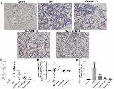
          Methods: Intratracheal injections of 3 mg/kg BLM were administered to induce pulmonary fibrosis in C57BL/6 mice. GHK was administered intraperitoneally at doses of 2.6, 26, and 260 μg/ml/day every other day from the 4th to the 21st day after BLM instillation. Three weeks after BLM instillation, pulmonary injury and pulmonary fibrosis was evaluated by the hematoxylin-eosin (HE) and Masson’s trichrome (MT) staining. Chronic inflammation index was used for the histological assessments by two pathologists blindly to each other. Tumor necrosis factor (TNF)-α and IL-6 levels in BALF and myeloperoxidase (MPO) activity in lung extracts were measured. For the pulmonary fibrosis evaluation, the fibrosis index calculated based on MT staining, collagen deposition and active TGF-β1 expression detected by ELISA, and the expression of TGF-β1, α-smooth muscle actin (SMA), fibronectin, MMP-9, and TIMP-1 by western blotting. The epithelial mesenchymal transition index, E-cadherin, and vimentin was also detected by western blot. The statistical analysis was performed by one-way ANOVA and the comparison between different groups were performed.

          Results: Treatment with GHK at all three doses reduced inflammatory cell infiltration and interstitial thickness and attenuated BLM-induced pulmonary fibrosis in mice. GHK treatment significantly improved collagen deposition, and MMP-9/TIMP-1 imbalances in lung tissue and also reduced TNF-α, IL-6 expression in bronchoalveolar lavage fluid (BALF) and MPO in lung extracts. Furthermore, GHK reversed BLM-induced increases in TGF-β1, p-Smad2, p-Smad-3 and insulin-like growth factor-1 (IGF-1) expression.

          Conclusion: GHK inhibits BLM-induced fibrosis progression, the inflammatory response and EMT via the TGF-β1/Smad 2/3 and IGF-1 pathway. Thus, GHK may be a potential treatment for pulmonary fibrosis.

          Related collections

          Most cited references38

          • Record: found
          • Abstract: found
          • Article: not found

          Natural inhibitor of transforming growth factor-beta protects against scarring in experimental kidney disease.

          The central pathological feature of human kidney disease that leads to kidney failure is the accumulation of extracellular matrix in glomeruli. Overexpression of transforming growth factor-beta (TGF-beta) underlies the accumulation of pathological matrix in experimental glomerulonephritis. Administration of an antibody raised against TGF-beta to glomerulonephritic rats suppresses glomerular matrix production and prevents matrix accumulation in the injured glomeruli. One of the matrix components induced by TGF-beta, the proteoglycan decorin, can bind TGF-beta and neutralize its biological activity, so decorin may be a natural regulator of TGF-beta (refs 3, 4). We tested whether decorin could antagonize the action of TGF-beta in vivo using the experimental glomerulonephritis model. We report here that administration of decorin inhibits the increased production of extracellular matrix and attenuates manifestations of disease, confirming our hypothesis. On the basis of our results, decorin may eventually prove to be clinically useful in diseases associated with overproduction of TGF-beta.
            Bookmark
            • Record: found
            • Abstract: found
            • Article: found
            Is Open Access

            Mechanisms of TGFβ-Induced Epithelial–Mesenchymal Transition

            Transitory phenotypic changes such as the epithelial–mesenchymal transition (EMT) help embryonic cells to generate migratory descendants that populate new sites and establish the distinct tissues in the developing embryo. The mesenchymal descendants of diverse epithelia also participate in the wound healing response of adult tissues, and facilitate the progression of cancer. EMT can be induced by several extracellular cues in the microenvironment of a given epithelial tissue. One such cue, transforming growth factor β (TGFβ), prominently induces EMT via a group of specific transcription factors. The potency of TGFβ is partly based on its ability to perform two parallel molecular functions, i.e. to induce the expression of growth factors, cytokines and chemokines, which sequentially and in a complementary manner help to establish and maintain the EMT, and to mediate signaling crosstalk with other developmental signaling pathways, thus promoting changes in cell differentiation. The molecules that are activated by TGFβ signaling or act as cooperating partners of this pathway are impossible to exhaust within a single coherent and contemporary report. Here, we present selected examples to illustrate the key principles of the circuits that control EMT under the influence of TGFβ.
              Bookmark
              • Record: found
              • Abstract: found
              • Article: not found

              Smad3 mediates transforming growth factor-beta-induced alpha-smooth muscle actin expression.

              Transforming growth factor-beta (TGF-beta)-induced alpha-smooth muscle actin (ASMA) expression is a key indicator of myofibroblast differentiation from fibroblasts. Recent studies suggest that a TGF-beta control element is important in the regulation of the ASMA gene promoter by TGF-beta. In this study, the role of Smad3, a key component of the Smad pathway that mediates TGF-beta signaling in regulation of ASMA gene expression, is investigated. All members of the Smad family were expressed in rat lung fibroblasts, and Smad3 expression was elevated upon TGF-beta 1 treatment. Transfection with a Smad3-expressing plasmid markedly increased Smad3 and ASMA protein expression, whereas transfection with an antisense Smad3 plasmid suppressed Smad3 and ASMA expression. Similar effects were noted when the cloned rat ASMA promoter-luciferase reporter gene construct was used to monitor transcriptional activation of the ASMA gene. Electrophoretic mobility shift assays and DNA affinity precipitation indicated Smad3 binding to at least two regions of the promoter containing CAGA motifs, termed Smad3-binding elements (SBEs). Mutation of one of the SBEs decreased promoter activity significantly, indicative of a functional role for this SBE. Taken together, these findings suggest a role for Smad3 in TGF-beta regulation of ASMA gene expression in myofibroblast differentiation.
                Bookmark

                Author and article information

                Contributors
                Journal
                Front Pharmacol
                Front Pharmacol
                Front. Pharmacol.
                Frontiers in Pharmacology
                Frontiers Media S.A.
                1663-9812
                12 December 2017
                2017
                : 8
                : 904
                Affiliations
                [1] 1Department of Respiratory Medicine, Shengjing Hospital of China Medical University , Shenyang, China
                [2] 2Department of Respiratory and Critical Care Medicine, The First Hospital of China Medical University , Shenyang, China
                Author notes

                Edited by: Alfonso Pompella, University of Pisa, Italy

                Reviewed by: Claudio Sorio, University of Verona, Italy; Amrita Dosanjh, Pediatric Respiratory Medical Group, United States

                *Correspondence: Gang Hou, hougangcmu@ 123456163.com

                This article was submitted to Experimental Pharmacology and Drug Discovery, a section of the journal Frontiers in Pharmacology

                Article
                10.3389/fphar.2017.00904
                5733019
                29311918
                366f82c4-249f-405c-bc38-06fec6735a16
                Copyright © 2017 Zhou, Wang, Wang, Liu, Zhang, Yin, Wang, Kang and Hou.

                This is an open-access article distributed under the terms of the Creative Commons Attribution License (CC BY). The use, distribution or reproduction in other forums is permitted, provided the original author(s) or licensor are credited and that the original publication in this journal is cited, in accordance with accepted academic practice. No use, distribution or reproduction is permitted which does not comply with these terms.

                History
                : 03 October 2017
                : 28 November 2017
                Page count
                Figures: 8, Tables: 1, Equations: 0, References: 44, Pages: 12, Words: 0
                Funding
                Funded by: Department of Education of Liaoning Province 10.13039/501100007620
                Award ID: LT2013015
                Categories
                Pharmacology
                Original Research

                Pharmacology & Pharmaceutical medicine
                ghk,tgf-β1,pulmonary fibrosis,epithelial-to-mesenchymal transition,smad,collagen

                Comments

                Comment on this article