1
views
0
recommends
+1 Recommend
0 collections
    0
    shares
      • Record: found
      • Abstract: found
      • Article: found
      Is Open Access

      Development and characterization of experimental ZnO cement containing niobophosphate bioactive glass as filling temporary material

      research-article

      Read this article at

      Bookmark
          There is no author summary for this article yet. Authors can add summaries to their articles on ScienceOpen to make them more accessible to a non-specialist audience.

          Abstract

          Aims

          The aim of this study was to develop and characterize a temporary restorative material based on a zinc oxide matrix containing niobophosphate bioactive glass (NbG) for the caries-affected dentin treatment.

          Material and methods

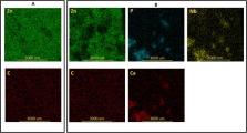
          NbG was added to a ZnO 2 matrix in different concentrations (wt%). EDS-SEM, ATR-FTIR and XRD analyses were performed to characterize the cement. Calcium release was evaluated in TRIS solution after 1, 7 and 14 days by colorimetric method (A 650). Compressive strengths and setting times were performed to analyze mechanical properties.

          Results

          EDS spectra confirmed the presence of Ca, P and Nb in the groups containing NbG. EDS mapping exhibit the ZnO 2 homogeneous distribution, and NbG immersed in this matrix. Peaks suggesting interaction between matrix and NbG were not detected in Ftir spectra. Calcium releasing showed to be time-dependent for experimental groups containing 10, 20, 30 and 40%. The NbG incorporation progressively increased the compressive strength values in the experimental groups. NbG incorporation seemed to influence the ZnO 2 matrix early setting reaction. No statistical difference was observed in the final setting time.

          Conclusion

          The addition of NbG particles into zinc oxide matrix could work as a mechanical reinforcement. It is suggested that the calcium released by the cement containing at least 10% NbG could induce apatite formation.

          Related collections

          Most cited references47

          • Record: found
          • Abstract: found
          • Article: not found

          The story of Bioglass.

          Historically the function of biomaterials has been to replace diseased or damaged tissues. First generation biomaterials were selected to be as bio-inert as possible and thereby minimize formation of scar tissue at the interface with host tissues. Bioactive glasses were discovered in 1969 and provided for the first time an alternative; second generation, interfacial bonding of an implant with host tissues. Tissue regeneration and repair using the gene activation properties of Bioglass provide a third generation of biomaterials. This article reviews the 40 year history of the development of bioactive glasses, with emphasis on the first composition, 45S5 Bioglass, that has been in clinical use since 1985. The steps of discovery, characterization, in vivo and in vitro evaluation, clinical studies and product development are summarized along with the technology transfer processes.
            Bookmark
            • Record: found
            • Abstract: not found
            • Article: not found

            Bioceramics: From Concept to Clinic

              Bookmark
              • Record: found
              • Abstract: found
              • Article: not found

              Regenerating bone with bioactive glass scaffolds: A review of in vivo studies in bone defect models.

              Large bone defects resulting from fractures and disease are a medical concern, being often unable to heal spontaneously by the body's repair mechanisms. Bone tissue engineering (BTE) is a promising approach for treating bone defects through providing a template to guide osseous regeneration. 3D scaffolds with microstructure mimicking host bone are necessary in common BTE strategies. Bioactive glasses (BGs) attract researchers' attention as BTE scaffolds as they are osteoconductive and osteoinductive in certain formulations. In vivo animal models allow understanding and evaluation of materials' performance in the complex physiological environment, being an inevitable step before clinical trials. The aim of this paper is to review for the first time published research investigating the in vivo osseous regenerative capacity of 3D BG scaffolds in bone defect animal models, to better understand and evaluate the progress and future outlook of the use of such scaffolds in BTE. The literature analysis reveals that the regenerative capacity of BG scaffolds depends on several factors; including BG composition, fabrication method, scaffold microstructure and pore characteristics, in addition to scaffold pretreatment and whether or not the scaffolds are loaded with growth factors. In addition, animal species selected, defect size and implantation time affect the scaffold in vivo behavior and outcomes. The review of the literature also makes clear the difficulty encountered to compare different types of bioactive glass scaffolds in their bone forming ability. Even considering such limitations of the current state-of-the-art, results generated from animal bone defect models provide an essential source of information to guide the design of BG scaffolds in future.
                Bookmark

                Author and article information

                Contributors
                Journal
                Saudi Dent J
                Saudi Dent J
                The Saudi Dental Journal
                Elsevier
                1013-9052
                1658-3558
                26 March 2021
                December 2021
                26 March 2021
                : 33
                : 8
                : 1111-1118
                Affiliations
                [a ]Piracicaba Dental School, Department of Restorative Dentistry, University of Campinas (UNICAMP), Limeira Av. 901, Piracicaba, São Paulo, Brazil
                [b ]General Dentistry Department, Division of Biomedical Materials, East Carolina University School of Dental Medicine, Greenville, NC, USA
                [c ]Department of Restorative Dentistry, School of Dentistry, University Ceuma (UNICEUMA), Josué Montello, 01, Renascença II, 65075120 São Luis, Maranhão, Brazil
                [d ]School of Dentistry, Discipline of Dental Materials, University Federal of Maranhão (UFMA), Campus Universitário do Bacanga, Av. dos Portugueses, 65085680 São Luis, MA, Brazil
                Author notes
                [* ]Corresponding author at: University Federal of Maranhão (UFMA), Av. dos Portugueses, 1966, 65085-680 São Luís, MA, Brazil. bauer@ 123456ufma.br
                Article
                S1013-9052(21)00036-5
                10.1016/j.sdentj.2021.03.009
                8665171
                34938056
                38ea96d4-e3a9-46e2-b9a7-282b6c00e453
                © 2021 The Authors

                This is an open access article under the CC BY-NC-ND license (http://creativecommons.org/licenses/by-nc-nd/4.0/).

                History
                : 30 June 2020
                : 9 March 2021
                : 10 March 2021
                Categories
                Original Article

                bioactive glass,bioactivity,ions release,restorative cement

                Comments

                Comment on this article