20
views
0
recommends
+1 Recommend
0 collections
    0
    shares
      • Record: found
      • Abstract: found
      • Article: found
      Is Open Access

      Effects of exosome-like vesicles on cumulus expansion in pigs in vitro

      research-article

      Read this article at

      Bookmark
          There is no author summary for this article yet. Authors can add summaries to their articles on ScienceOpen to make them more accessible to a non-specialist audience.

          Abstract

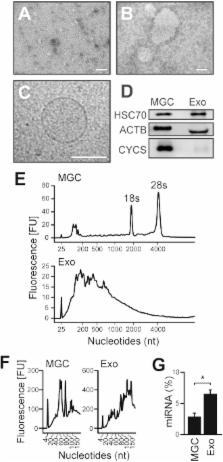
          Cell-secreted vesicles, such as exosomes, have recently been recognized as mediators of cell communication. A recent study in cattle showed the involvement of exosome-like vesicles in the control of cumulus expansion, a prerequisite process for normal ovulation; however, whether this is the case in other mammalian species is not known. Therefore, this study aimed to examine the presence of exosome-like vesicles in ovarian follicles and their effects on cumulus expansion in vitro in pigs. The presence of exosome-like vesicles in porcine follicular fluid (pFF) was confirmed by transmission electron microscopic observation, the detection of marker proteins, and RNA profiles specific to exosomes. Fluorescently labeled exosome-like vesicles isolated from pFF were incorporated into both cumulus and mural granulosa cells in vitro. Exosome-like vesicles were not capable of inducing cumulus expansion to a degree comparable to that induced by follicle-stimulating hormone (FSH). Moreover, exosome-like vesicles had no significant effects on the expression levels of transcripts required for the normal expansion process (HAS2, TNFAIP6, and PTGS2). Interestingly, FSH-induced expression of HAS2 and TNFAIP6 mRNA, but not of PTGS2 mRNA, was significantly increased by the presence of exosome-like vesicles; however, the degree of FSH-induced expansion was not affected. In addition, porcine exosome-like vesicles had no significant effects on the expansion of mouse cumulus-oocyte complexes. Collectively, the present results suggest that exosome-like vesicles are present in pFF, but they are not efficient in inducing cumulus expansion in pigs.

          Related collections

          Most cited references39

          • Record: found
          • Abstract: found
          • Article: not found

          Electron microscopic evidence for externalization of the transferrin receptor in vesicular form in sheep reticulocytes

          Using ferritin-labeled protein A and colloidal gold-labeled anti-rabbit IgG, the fate of the sheep transferrin receptor has been followed microscopically during reticulocyte maturation in vitro. After a few minutes of incubation at 37 degrees C, the receptor is found on the cell surface or in simple vesicles of 100-200 nm, in which the receptor appears to line the limiting membrane of the vesicles. With time (60 min or longer), large multivesicular elements (MVEs) appear whose diameter may reach 1-1.5 micron. Inside these large MVEs are round bodies of approximately 50-nm diam that bear the receptor at their external surfaces. The limiting membrane of the large MVEs is relatively free from receptor. When the large MVEs fuse with the plasma membrane, their contents, the 50-nm bodies, are released into the medium. The 50-nm bodies appear to arise by budding from the limiting membrane of the intracellular vesicles. Removal of surface receptor with pronase does not prevent exocytosis of internalized receptor. It is proposed that the exocytosis of the approximately 50-nm bodies represents the mechanism by which the transferrin receptor is shed during reticulocyte maturation.
            Bookmark
            • Record: found
            • Abstract: found
            • Article: not found

            EGF-like growth factors as mediators of LH action in the ovulatory follicle.

            Before ovulation in mammals, a cascade of events resembling an inflammatory and/or tissue remodeling process is triggered by luteinizing hormone (LH) in the ovarian follicle. Many LH effects, however, are thought to be indirect because of the restricted expression of its receptor. Here, we demonstrate that LH stimulation induces the transient and sequential expression of the epidermal growth factor (EGF) family members amphiregulin, epiregulin, and beta-cellulin. Incubation of follicles with these growth factors recapitulates the morphological and biochemical events triggered by LH, including cumulus expansion and oocyte maturation. Thus, these EGF-related growth factors are paracrine mediators that propagate the LH signal throughout the follicle.
              Bookmark
              • Record: found
              • Abstract: found
              • Article: not found

              Cell-secreted vesicles in equine ovarian follicular fluid contain miRNAs and proteins: a possible new form of cell communication within the ovarian follicle.

              Proper cell communication within the ovarian follicle is critical for the growth and maturation of a healthy oocyte that can be fertilized and develop into an embryo. Cell communication within the follicle involves many signaling molecules and is affected by maternal age. Recent studies indicate that cell communication can be mediated through secretion and uptake of small membrane-enclosed vesicles. The goals of this study were to 1) identify cell-secreted vesicles (microvesicles and exosomes) containing miRNAs and proteins within ovarian follicular fluid and 2) determine if miRNA level differs in exosomes isolated from follicular fluid in young compared to old mares. We demonstrate the presence of vesicles resembling microvesicles and exosomes in ovarian follicular fluid using transmission electron microscopy and CD63-positive and RNA containing vesicles using flow cytometry. Moreover, proteomics analysis reveals that follicular fluid-isolated exosomes contain both known exosomal proteins and proteins not previously reported in isolated exosomes. MicroRNAs were detected in microvesicle and exosomes preparations isolated from follicular fluid by real-time PCR analysis. Uptake of fluorescent-labeled microvesicles by granulosa cells was examined using in vitro and in vivo approaches. MicroRNA expression profiling reveals that miRNAs in microvesicle and exosome preparations isolated from follicular fluid also are present within surrounding granulosa and cumulus cells. These studies revealed that cell communication within the mammalian ovarian follicle may involve transfer of bioactive material by microvesicles and exosomes. Finally, miRNAs present in exosomes from ovarian follicular fluid varied with the age of the mare, and a number of different miRNAs were detected in young vs. old mare follicular fluid.
                Bookmark

                Author and article information

                Journal
                J Reprod Dev
                J. Reprod. Dev
                JRD
                The Journal of Reproduction and Development
                The Society for Reproduction and Development
                0916-8818
                1348-4400
                05 February 2017
                February 2017
                : 63
                : 1
                : 51-58
                Affiliations
                [1) ]Laboratory of Applied Genetics, Department of Animal Resource Sciences, Graduate School of Agriculture and Life Sciences, The University of Tokyo, Tokyo 113-8657, Japan
                [2) ]Biomimetic Materials Science Laboratory, Graduate School of Materials Science, Nara Institute of Science and Technology (NAIST), Nara 630-0192, Japan
                Author notes
                Correspondence: K Sugiura (e-mail: aks@ 123456mail.ecc.u-tokyo.ac.jp )
                Article
                2016-124
                10.1262/jrd.2016-124
                5320430
                28163264
                3b2fe2dd-3c0e-453b-b94f-c3c7de27752d
                ©2017 Society for Reproduction and Development

                This is an open-access article distributed under the terms of the Creative Commons Attribution Non-Commercial No Derivatives (by-nc-nd) License. (CC-BY-NC-ND 4.0: http://creativecommons.org/licenses/by-nc-nd/4.0/ )

                History
                : 17 August 2016
                : 30 September 2016
                Categories
                Original Article

                extracellular vesicles,granulosa cells,ovarian follicles,porcine follicular fluid

                Comments

                Comment on this article