0
views
0
recommends
+1 Recommend
0 collections
    0
    shares
      • Record: found
      • Abstract: found
      • Article: found
      Is Open Access

      Perspective: Structure determination of protein-ligand complexes at room temperature using X-ray diffraction approaches

      brief-report

      Read this article at

      Bookmark
          There is no author summary for this article yet. Authors can add summaries to their articles on ScienceOpen to make them more accessible to a non-specialist audience.

          Abstract

          The interaction between macromolecular proteins and small molecule ligands is an essential component of cellular function. Such ligands may include enzyme substrates, molecules involved in cellular signalling or pharmaceutical drugs. Together with biophysical techniques used to assess the thermodynamic and kinetic properties of ligand binding to proteins, methodology to determine high-resolution structures that enable atomic level interactions between protein and ligand(s) to be directly visualised is required. Whilst such structural approaches are well established with high throughput X-ray crystallography routinely used in the pharmaceutical sector, they provide only a static view of the complex. Recent advances in X-ray structural biology methods offer several new possibilities that can examine protein-ligand complexes at ambient temperature rather than under cryogenic conditions, enable transient binding sites and interactions to be characterised using time-resolved approaches and combine spectroscopic measurements from the same crystal that the structures themselves are determined. This Perspective reviews several recent developments in these areas and discusses new possibilities for applications of these advanced methodologies to transform our understanding of protein-ligand interactions.

          Related collections

          Most cited references53

          • Record: found
          • Abstract: found
          • Article: not found

          The potential and limitations of neutrons, electrons and X-rays for atomic resolution microscopy of unstained biological molecules.

          Radiation damage is the main problem which prevents the determination of the structure of a single biological macromolecule at atomic resolution using any kind of microscopy. This is true whether neutrons, electrons or X-rays are used as the illumination. For neutrons, the cross-section for nuclear capture and the associated energy deposition and radiation damage could be reduced by using samples that are fully deuterated and 15N-labelled and by using fast neutrons, but single molecule biological microscopy is still not feasible. For naturally occurring biological material, electrons at present provide the most information for a given amount of radiation damage. Using phase contrast electron microscopy on biological molecules and macromolecular assemblies of approximately 10(5) molecular weight and above, there is in theory enough information present in the image to allow determination of the position and orientation of individual particles: the application of averaging methods can then be used to provide an atomic resolution structure. The images of approximately 10,000 particles are required. Below 10(5) molecular weight, some kind of crystal or other geometrically ordered aggregate is necessary to provide a sufficiently high combined molecular weight to allow for the alignment. In practice, the present quality of the best images still falls short of that attainable in theory and this means that a greater number of particles must be averaged and that the molecular weight limitation is somewhat larger than the predicted limit. For X-rays, the amount of damage per useful elastic scattering event is several hundred times greater than for electrons at all wavelengths and energies and therefore the requirements on specimen size and number of particles are correspondingly larger. Because of the lack of sufficiently bright neutron sources in the foreseeable future, electron microscopy in practice provides the greatest potential for immediate progress.
            Bookmark
            • Record: found
            • Abstract: found
            • Article: found
            Is Open Access

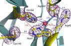
            Structural plasticity of SARS-CoV-2 3CL M pro active site cavity revealed by room temperature X-ray crystallography

            The COVID-19 disease caused by the SARS-CoV-2 coronavirus has become a pandemic health crisis. An attractive target for antiviral inhibitors is the main protease 3CL Mpro due to its essential role in processing the polyproteins translated from viral RNA. Here we report the room temperature X-ray structure of unliganded SARS-CoV-2 3CL Mpro, revealing the ligand-free structure of the active site and the conformation of the catalytic site cavity at near-physiological temperature. Comparison with previously reported low-temperature ligand-free and inhibitor-bound structures suggest that the room temperature structure may provide more relevant information at physiological temperatures for aiding in molecular docking studies.
              Bookmark
              • Record: found
              • Abstract: found
              • Article: not found

              Accessing protein conformational ensembles using room-temperature X-ray crystallography.

              Modern protein crystal structures are based nearly exclusively on X-ray data collected at cryogenic temperatures (generally 100 K). The cooling process is thought to introduce little bias in the functional interpretation of structural results, because cryogenic temperatures minimally perturb the overall protein backbone fold. In contrast, here we show that flash cooling biases previously hidden structural ensembles in protein crystals. By analyzing available data for 30 different proteins using new computational tools for electron-density sampling, model refinement, and molecular packing analysis, we found that crystal cryocooling remodels the conformational distributions of more than 35% of side chains and eliminates packing defects necessary for functional motions. In the signaling switch protein, H-Ras, an allosteric network consistent with fluctuations detected in solution by NMR was uncovered in the room-temperature, but not the cryogenic, electron-density maps. These results expose a bias in structural databases toward smaller, overpacked, and unrealistically unique models. Monitoring room-temperature conformational ensembles by X-ray crystallography can reveal motions crucial for catalysis, ligand binding, and allosteric regulation.
                Bookmark

                Author and article information

                Contributors
                Journal
                Front Mol Biosci
                Front Mol Biosci
                Front. Mol. Biosci.
                Frontiers in Molecular Biosciences
                Frontiers Media S.A.
                2296-889X
                23 January 2023
                2023
                : 10
                : 1113762
                Affiliations
                [1] 1 School of Life Sciences , University of Essex , Colchester, United Kingdom
                [2] 2 Diamond Light Source Ltd. , Harwell Science and Innovation Campus , Didcot, United Kingdom
                Author notes

                Edited by: Gourinath Samudrala, Jawaharlal Nehru University, India

                Reviewed by: Sudhaker Dharavath, National Cancer Institute at Frederick (NIH), United States

                *Correspondence: Michael A. Hough, michael.hough@ 123456diamond.ac.uk ; Jonathan A. R. Worrall, jworrall@ 123456essex.ac.uk

                This article was submitted to Structural Biology, a section of the journal Frontiers in Molecular Biosciences

                Article
                1113762
                10.3389/fmolb.2023.1113762
                9899996
                3b708f6f-c586-4135-b1e1-0ab704e6076a
                Copyright © 2023 Hough, Prischi and Worrall.

                This is an open-access article distributed under the terms of the Creative Commons Attribution License (CC BY). The use, distribution or reproduction in other forums is permitted, provided the original author(s) and the copyright owner(s) are credited and that the original publication in this journal is cited, in accordance with accepted academic practice. No use, distribution or reproduction is permitted which does not comply with these terms.

                History
                : 01 December 2022
                : 12 January 2023
                Funding
                Funded by: Biotechnology and Biological Sciences Research Council , doi 10.13039/501100000268;
                Award ID: BB/W001950/1 BB/01577X/1 BB/R021015/1
                Funded by: Leverhulme Trust , doi 10.13039/501100000275;
                Award ID: RPG-2014-355
                Categories
                Molecular Biosciences
                Perspective

                x-ray crystallography,time-resolved,ambient temperature,spectroscopy,protein-ligand complexes

                Comments

                Comment on this article